
Page 227 - IJB-8-4
P. 227
Neng, et al.
team proposed an in situ bioprinting method based on
this platform, used a non-planar slice algorithm for path
planning and design, and developed a contact probe as the
end execution of the robotic arm device. The probe can
realize the surface reconstruction of the defect, record the
penetration depth of the penetration point, and help plan
the path. Finally, the researchers used the printing method
to test anthropomorphic models to repair skull defects .
[49]
The team of Professor Xu Tao at Tsinghua University
proposed a concept of in situ bioprinting in vivo. They
developed a miniature in situ bioprinting robot that can
be mounted to an endoscope, which can enter the human
body for in situ printing . They use the miniature robot to
[50]
treat gastric wall injuries. This technology miniaturizes the
equipment and implants it into the human body for in situ
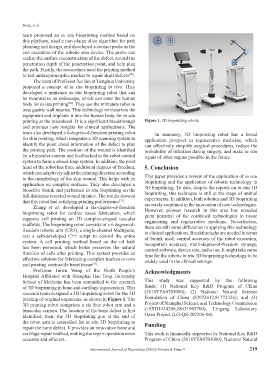
printing at the microlevel. It is a significant breakthrough Figure 1. 3D bioprinting robots.
and provides new insights for clinical applications. The
team also developed a 6-degree-of-freedom printing robot In summary, 3D bioprinting robot has a broad
for skin printing, which integrates a 3D scanning system to application prospect in regenerative medicine, which
identify the point cloud information of the defect to plan can effectively simplify surgical procedures, reduce the
the printing path. The position of the wound is identified probability of infection during surgery, and make in situ
by a binocular camera and feedbacked to the robot control repair of other organs possible in the future.
system to form a closed-loop system. In addition, the print
head of the robot has three additional degrees of freedom, 5. Conclusion
which can adaptively adjust the printing direction according
to the morphology of the skin wound. This helps with its This paper provides a review of the application of in situ
application on complex surfaces. They also developed a bioprinting and the application of robotic technology in
bioactive bioink and performed in situ bioprinting on the 3D bioprinting. To date, despite the reports on in situ 3D
full-thickness resected wound in mice. The results showed bioprinting, this technique is still at the stage of animal
experiments. In addition, both robotics and 3D bioprinting
that this robot had satisfying printing performance . are rarely combined in the innovation of new technologies.
[51]
Zhang et al. developed a six-degree-of-freedom
bioprinting robot for cardiac tissue fabrication, which However, pioneer research in this area has revealed
great potential of the combined technologies in tissue
supports cell printing on 3D complex-shaped vascular engineering and regenerative medicine. Nevertheless,
scaffolds. The bioprinting robot consists of a 6 degree-of- there are still some difficulties in applying this technology
freedom robotic arm (UR3), a single-channel Multipette, to clinical applications. Breakthroughs are needed in terms
and a self-developed C++ script to control the entire of bioink used, control accuracy of the robot execution,
system. A cell printing method based on the oil bath recognition accuracy, multi-degree-of-freedom synergy,
has been proposed, which better preserves the natural control software, device size, and so on. It might take some
function of cells after printing. This system provides an time for the robotic in situ 3D bioprinting technology to be
effective solution for fabricating complex trachea in vitro widely used in the clinical settings.
and printing contractile heart tissue .
[52]
Professor Jinwu Wang of the Ninth People’s Acknowledgments
Hospital Affiliated with Shanghai Jiao Tong University
School of Medicine has been committed to the research This study was supported by the following
of 3D bioprinting in bone and cartilage regeneration. This funds: (1) National Key R&D Program of China
research team designed a 3D bioprinting robot for the 3D (2018YFA0703000); (2) National Natural Science
printing of original organisms, as shown in Figure 1. The Foundation of China (82072412,81772326); and (3)
3D printing robot comprises a six free robot arm and a Project of Shanghai Science and Technology Commission
binocular camera. The location of the bone defect is first (19XD1434200,18431903700); Lingang Laboratory
identified; then, the 3D bioprinting gun at the end of Open Project (LG-QS-202206-04)
the robot arm is controlled for in situ 3D bioprinting to Funding
repair the bone defect. It provides an innovative bone and
cartilage repair method, making the repair operation more This work is financially supported by National Key R&D
accurate and efficient. Program of China (2018YFA0703000), National Natural
International Journal of Bioprinting (2022)–Volume 8, Issue 4 219