
Page 63 - IJB-8-4
P. 63
Fang, et al.
chips; however, the number of cells on the chips was in the area-to-volume ratio and shear stresses were credited for
order of 300 µm > 500 µm > 700 µm. The high surface- high cell growth in the chips with narrower channels.
Drug metabolism was investigated by seeding 7-ethoxy-
4-trifluoromethyl coumarin (EFC) solution into the cell-
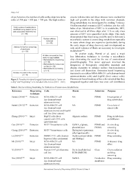
Total number of publications laden chips. Metabolism of EFC to its metabolite, HFC,
identified using search terms
and associated MeSH terms was observed in all three chips after 12 h as only trace
(n = 98) amounts of EFC were quantified on the chips. This study
demonstrated that bioprinting could be used to fabricate a
Review articles microfluidic system to assess drug metabolism especially
removed
(n = 35) in cases where drugs are expensive (particularly during
the early stages of drug discovery and development) as
Articles for further screening only small volumes of fluids are necessary to investigate
of abstracts
(n = 63) metabolism [123] .
In another study, Hamid et al. used a mask
Articles removed as they less fabrication technique to develop a microfluidic
were not appropriate
• Not related to bioprinting chip eliminating the need for the use of conventional
(n = 38) photolithography. This novel approach involved the
• Not related to tumour-on- integration of biologically compatible materials and
a-chip (n = 12)
• Not related to screening plasma chemistry to enhance surface functionalization
(n = 4) and direct cell deposition. Extrusion-based bioprinting
Articles included in the review was used to co-culture MDA-MB-231 cells (human breast
(n = 9)
adenocarcinoma cells) and HepG2 (liver cancer cells).
Figure 5. Flowchart for identifying publications related to “tumor-on- Fluorescent-based tracking of the cells indicated that they
a-chip” and “bioprinting” to be included in the review (2013 – 2021). integrated together and there was even cell distribution
Table 1. Studies utilizing bioprinting for fabrication of tumor-on-a-chip platforms.
Reference Bioprinting Cells Bioink Substrate Purpose
technique
Hamid (2014) [117] Extrusion MDA-MB-231 cell NA PDMS Investigation of
line (human breast drug metabolism
adenocarcinoma)
Hamid (2015) [118] Extrusion MDA-MB-231 cell NA PDMS Co-culture of
line (human breast cancer cells
adenocarcinoma) and HepG2
cell line (liver cancer)
Zhang (2016) [119] Inkjet HepG2 cells (liver Alginate sodium PDMS Drug metabolism
cancer) and U251 cells and diffusion
(glioblastoma)
Cao (2019) [13] Extrusion MCF-7 breast tumor cells GelMA, alginate, PDMS and In vitro drug
photoinitiator PMMA screening
Cheng (2019) [120] Extrusion MCF-7 breast tumor cells Hydrophobic petroleum In vitro drug
jelly-liquid paraffin screening
Li (2019) [121] Extrusion SMMC-7721 cells (human Hydroxypropyl chitin PDMS Drug screening
hepatocellular carcinoma) and Matrigel
Mi (2019) [122] Inkjet MDA-MB-231 cell NA PDMS Anticancer drug
line (human breast effect
adenocarcinoma)
Yi (2019) [123] Extrusion Human U-87 glioblastoma Brain decellularized Glass Chemoradiation
cell line ECM (BdECM) and drug
screening
Xie (2020) [124] Inkjet MDA-MB-231 cell GelMA Conductive Drug screening
line (human breast membrane
adenocarcinoma)
International Journal of Bioprinting (2022)–Volume 8, Issue 4 55