
Page 60 - IJB-8-4
P. 60
3D Bioprinting for Anticancer Drug Screening
densities. This method has been used in the regeneration
of skin and cartilage. Human ear and sheep meniscus have
been inkjet-bioprinted using nanocellulose bioink [92,93] .
The deposition of material extruded through a
nozzle in a well-defined continuous stream is the basis
for extrusion-based bioprinting. It permits deposition
of materials in higher viscosity compared to inkjet
bioprinting, making it suited for rebuilding highly cellular
tissues. However, the shear stresses created can lead to
cellular damage. To maintain cell stability and survival,
strict control of the quantity of biomaterial, pressure, and
nozzle diameter is required [18,82] . Aortic valve conduits,
vascular grafts, and cartilage structures have all been
printed using this technology [94-96] .
LAB is a relatively new technology in which a
laser pulse is focused on the donor ribbon and converted
into a shockwave to activate the bioink underneath .
[81]
The high resolution of LAB (~10 µm) allows it to be
used to make structures of native tissues at or near the
scale of a single cell and its contactless and nozzle-free
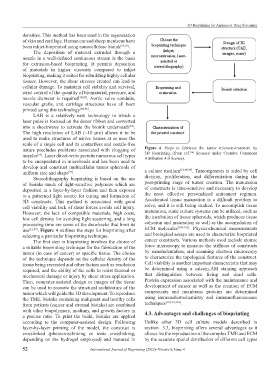
nature precludes problems associated with clogging of Figure 4. Steps to fabricate the tumor microenvironment by
3D bioprinting (from ref. licensed under Creative Commons
[16]
nozzles . Laser direct-write permits numerous cell types Attribution 4.0 license).
[92]
to be encapsulated in microbeads and has been used to
develop and construct multicellular tumor spheroids of [18,98,99]
uniform size and shape . a culture medium . Tumorigenesis is aided by cell
[97]
Stereolithography bioprinting is based on the use division, proliferation, and differentiation during the
of bioinks made of light-sensitive polymers which are post-printing stage of tumor creation. The maturation
deposited in a layer-by-layer fashion and then exposed of constructs is time-sensitive and necessary to develop
to a patterned light source for curing and formation of the most effective personalized anticancer regimen.
3D constructs. This method is associated with good Accelerated tissue maturation is a difficult problem to
cell viability and lack of shear forces avoids cell injury. solve, and it is still being studied. To accomplish tissue
However, the lack of compatible materials, high costs, maturation, static culture systems can be utilized, such as
low cell density for avoiding light scattering, and a long the incubation of tissue spheroids, which produces tissue
processing time are some of the drawbacks that limit its cohesion and maturation as well as the accumulation of
[100-102]
use [12,87] . Figure 4 outlines the steps for bioprinting after ECM molecules . Physicochemical measurements
selecting a particular bioprinting technique. and biological assays are used to characterize bioprinted
The first step in bioprinting involves the choice of cancer constructs. Various methods used include atomic
a suitable bioprinting technique for the fabrication of the force microscopy to measure the stiffness of constructs
tumor (in case of cancer) or specific tissue. The choice by nanoindentation, and scanning electron microscopy
of the technique depends on the cellular density of the to characterize the topological features of the construct.
tissue being recreated and other factors such as resolution Cell viability is another important characteristic that may
required, and the ability of the cells to resist thermal or be determined using a calcein-AM staining approach
mechanical damage or injury by shear stress application. that distinguishes between living and dead cells.
Then, computer-assisted design or images of the tissue Protein expression associated with the maintenance and
can be used to recreate the structural architecture of the development of cancer as well as the creation of ECM
tumor which will guide the 3D development. To reproduce components and membrane proteins are determined
the TME, bioinks containing malignant and healthy cells using immunohistochemistry and immunofluorescence
from patients (cancer and stromal bioinks) are combined techniques [18,103,104] .
with other biopolymers, medium, and growth factors in 4.3. Advantages and challenges of bioprinting
a precise ratio. To print the build, bioinks are applied
according to the computer-assisted design. Following Unlike other 3D cell culture models described in
layer-by-layer printing of the model, the construct is section 3.3, bioprinting offers several advantages as it
crosslinked (photocrosslinking or ionic crosslinking, allows for the reproduction of the complex TME and ECM
depending on the hydrogel employed) and matured in by the accurate spatial distribution of different cell types
52 International Journal of Bioprinting (2022)–Volume 8, Issue 4