
Page 57 - IJB-8-4
P. 57
Fang, et al.
Several limitations associated with 2D cell culture
models have led to the development and use of in vivo
animal models and 3D models that can closely mimic the
physical, geometrical, biochemical, and genetic profiles
of the TME.
3.2. In vivo animal models
In vivo investigations in animal models are most
often the next logical step following successful
in vitro determination of the efficacy of potential drug
candidates. Although in vivo models provide essential
information on tumor growth and progression, they
suffer from several limitations, such as high cost,
large variations observed between the animals used,
inter-species differences, and ethical considerations
relating to animal use. Because of the variations in
gene expression, protein expression, and soluble
molecules (cytokines, growth factors, etc.) that are
crucial for the investigation of cancer progression,
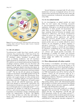
Figure 1. Various in vitro cancer models are used in anticancer drug animal models such as rat models utilized in cancer
screening (from reference [19] licensed under Creative Commons research are not indicative of human responses to
Attribution 4.0 license) anticancer treatments [66-68] . Despite their limitations,
animal models continue to remain the gold standard
for anticancer compounds due to their physiological
3.1. 2D cell cultures similarity to humans. The large gap between 2D
Two-dimensional models have been typically used for cell cultures and animal models necessitates the
anticancer drug screening due to their ease of use, low development of suitable 3D models that eliminate
cost, and availability of functional assays . Traditional species differences in drug response observed during
[60]
2D monolayer culture, monolayer co-cultures, cells animal testing and enables the recreation of the
grown on floating membranes, and cell monolayer complex TME.
sandwiched between membranes are the most often used 3.3. Three-dimensional cell culture models
2D models in cancer research and screening. Despite their
advantages related to usability and cost, 2D model cell The advances in microfluidics, cell biology, and tissue
cultures lack the ability to accurately identify potential engineering have paved the way for the creation of 3D
anticancer compounds aimed at treating solid tumors. In cell culture models that can mimic the pathophysiology of
cancer patients, drugs that have demonstrated substantial tumor cells and their responses to anticancer treatments.
performance in 2D in vitro models have not always worked Correct cell polarization, original form, genetic profile,
as well . The fact that 2D cell cultures are frequently and TME heterogeneity may all be obtained using 3D
[61]
[12]
produced as monolayers, which differ greatly from the 3D models . Tumors are 3D formations made up of a
architecture of tumors, explain this phenomenon. Drug, heterogeneous distribution of various cells buried in
nutrients, and oxygen exposure are often uniform across stromal tissue; therefore, 3D models can better depict the
all cells in a monolayer whereas inner tumor cells have TME and cellular behavior .
[19]
limited drug exposure . Furthermore, 2D models cannot To create realistic human models for chemotherapeutic
[62]
capture cell-cell and cell-ECM interactions, and they drug testing, primary cancer cells obtained from patient
suffer from damage to tissue-specific construction, and tumor biopsies or cell lines can be employed. The different
biochemical and mechanical cues caused by the removal 3D cell cultures include spheroids, organoids, scaffolds/
of secreted biomolecules by media replacement [63-65] . Two- hydrogels, and 3D-bioprinted constructs.
dimensional cell cultures are made up of immortalized Spheroids are micro-sized, single-cell, or multicell
cell lines, and recurrent passages of cell lines can result aggregates that represent the 3D architecture of solid
in genotypic and phenotypic changes, obstructing proper tumors and they can demonstrate cell-cell and cell-
growth and responsiveness to stimuli. Cell supports ECM interactions, unlike 2D cell culture systems [69,70] .
in Petri dishes or flasks are stiffer than soft tissues of Spheroids can form intrinsic metabolic gradients (oxygen,
tumors, which affects gene expression profiles and drug nutrients, by-products, and metabolites) that lead to the
sensitivity . establishment of heterogenous structures composed of a
[66]
International Journal of Bioprinting (2022)–Volume 8, Issue 4 49