
Page 59 - IJB-8-4
P. 59
Fang, et al.
and appropriate MesH terms. A clear upward trend in Cancer bioinks consist of patient-derived cancer
research related to 3D bioprinting technology is evident. cells (primary cancer cells, cancer stem cells, circulating
Several review papers on the evolution of cell cultures tumor cells, and CAFs) mixed with a biopolymer
for cancer drug screening, organ-on-chip platforms, and (GelMA, a methacrylate form of gelatin, alginate,
bioprinting have also been published. hyaluronic acid, or collagen), growth agents, and
nutrition. Matrigel, a gelatinous ECM protein generated
4.1. Bioinks by mouse sarcoma cells, is commonly utilized to provide
Bioinks are important tools for the fabrication of artificial the ideal environment for cancer cell proliferation and
living tissue constructs that can mimic all properties of carcinogenesis, as well as to imitate the morphological
native tissues through 3D printing technologies. Bioinks are properties of in vivo tumors [18,89] .
the building blocks of bioprinted constructions, consisting Stromal bioinks contain healthy stromal cells such
of a biocompatible hydrogel in which the cells of interest as endothelial cells, mesenchymal/hematopoietic stem
as well as nutrition and growth factors are embedded . cells, fibroblasts, and other tissue cells. Patient-derived
[81]
Biocompatibility, predictable gelation, capacity to imitate primary cells or BioBank primary cell lines should be
structural, physicochemical, rheological and biological used to mimic the microenvironment along with natural
features of the ECM, amenability to scale-up, production and synthetic polymers. Furthermore, endothelial cells or
under good manufacturing principles, and minimal endothelial progenitor cells should also be incorporated
batch-to-batch variability are all desired qualities of the with angiogenic factors to recapitulate tumor blood
hydrogels [18,87] . They can be composed of only cells, but vessel features, such as extensive branching and leaky
usually, a carrier substance such as a natural or synthetic vasculature [18,90,91] .
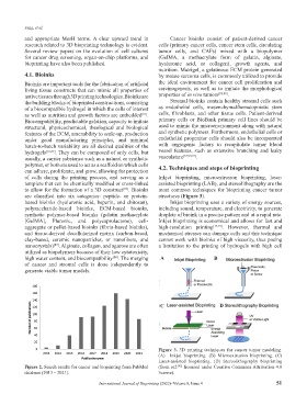
polymer, or both are used to act as a scaffold on which cells 4.2. Techniques and steps of bioprinting
can adhere, proliferate, and grow, allowing for protection
of cells during the printing process, and serving as a Inkjet bioprinting, microextrusion bioprinting, laser-
template that can be chemically modified or cross-linked assisted bioprinting (LAB), and stereolithography are the
to allow for the formation of a 3D construct . Bioinks most common techniques for bioprinting cancer tumor
[87]
are classified into six categories: peptide- or protein- structures (Figure 3).
based bioinks (hyaluronic acid, heparin, and chitosan), Inkjet bioprinting uses a variety of energy sources,
polysaccharide-based bioinks, ECM-based bioinks, including sound, temperature, and electricity, to generate
synthetic polymer-based bioinks (gelatin methacrylate droplets of bioink in a precise pattern and at a rapid rate.
[GelMA], Pluronic, and polycaprolactone), cell- Inkjet bioprinting is economical and allows for fast and
aggregate or pellet-based bioinks (fibrin-based bioinks), high-resolution printing [12,19] . However, thermal and
and tissue-derived decellularized matrix (carbon-based, mechanical stresses can damage cells and this technique
clay-based, ceramic nanoparticles, or nanofibers, and cannot work with bioinks of high viscosity, thus posing
nanocrystals) . Alginate, collagen, and agarose are often a limitation to the printing of hydrogels with high cell
[87]
utilized as biopolymers because of their low cytotoxicity,
high water content, and biocompatibility . The merging A B
[88]
of cancer and stromal cells is done independently to
generate viable tumor models.
C D
Figure 3. 3D printing techniques for cancer tumor modeling.
(A) Inkjet bioprinting. (B) Microextrusion bioprinting. (C)
Laser-assisted bioprinting. (D) Stereolithography bioprinting
Figure 2. Search results for cancer and bioprinting from PubMed (from ref. [92] licensed under Creative Commons Attribution 4.0
database (2013 – 2021). license).
International Journal of Bioprinting (2022)–Volume 8, Issue 4 51