
Page 189 - IJB-9-3
P. 189
International Journal of Bioprinting Chitin/gelatin/PVA scaffolds
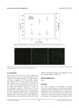
Figure 6. Biocompatibility assessment through the exposition of the scaffold to HS27 cells at 24, 48 and 72 h of culture.
Figure 7. Images taken following the live/dead cell viability assay at 1, 3, and 7 days of culture after the direct exposure of the scaffolds. Live and dead cells
are shown in green and red, respectively. Scale bar represents 50 µm.
4. Conclusion adhesion and growth. Therefore, cell viability was tested
and biocompatibility was confirmed.
Fly fruit pupae were found to be good candidates for
chitin extraction, as proven by the high extraction yield
obtained. Additionally, the purity of the chitin obtained Acknowledgments
was high, as suggested by FTIR and XRD analyses. None.
This chitin was used to prepare gelatin-based inks for
3DP, while ES inks were prepared with gelatin and
PVA. Both inks had shear thinning behavior, required Funding
for additive manufacturing processes, as shown by This work was supported by the Basque Government
the rheological assessment. Furthermore, 3DP inks through the research groups of the Basque university
exhibited good mucoadhesion and textural properties system (IT1658-22) and through Elkartek Program (KK-
for tissue engineering applications. It is worth noting 2022/00019). Work at Biodonostia was supported by
the good replicability of the scaffolds manufactured by Project PI19/01621, funded by Instituto de Salud Carlos III
combining 3DP and ES. Resulting scaffolds showed an (ISCIII) and co-funded by the European Union. J.U. thanks
interconnected pore architecture, favorable for cellular the University of the Basque Country (ESPDOC21/74).
Volume 9 Issue 3 (2023) 181 https://doi.org/10.18063/ijb.701