Page 188 - IJB-9-3
P. 188
International Journal of Bioprinting                                          Chitin/gelatin/PVA scaffolds


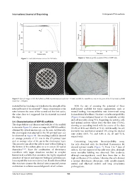
                   Figure 3. Load-deformation curves for 3DP inks.   Figure 4. Textural profile analysis (TPA) of 3DP inks.


            Figure 5. Optical images of (A) the hybrid scaffold macrostructure (scale bar = 4 mm) and (B) the nanofibrous mat on the pore of the 3D-printed scaffold
            (scale bar = 200 µm).
            material before breaking and it indicates the strength of the   With the aim of assessing the potential of these
            internal bonds of the material . Since cohesiveness is the   multilayered scaffolds for tissue regeneration, such as
                                    [59]
            ratio between the areas of the second and the first cycles,   wound  healing,  biocompatibility  was  determined  using
            the value close to 1 suggested that the material recovered   human dermal fibroblasts. The short-term biocompatibility
            the shape.                                         (Figure 6) was evaluated based on the metabolic activity
                                                               and cell mortality along 72 h. Regarding the activity, cells
            3.4. Characterization of 3DP-ES scaffolds          had optimal activity values from the first date (75.0%),
            The shape fidelity and dimensional stability of the scaffold   showing an ascending trend with the course of the days
            is shown in Figure 5A, where an image of a 3DP-ES scaffold,   (91.8% at 48 h and 106.8% at 72 h). Additionally, the cell
            obtained by optical microscopy, can be seen. Additionally,   mortality was maintained around 10% along the studied
            the electrospun mat attached to the 3D-printed layer can   time points (10.3, 7.6, and 8.4% at 24, 48 and 72 h,
            be observed in Figure 5B. The resulting scaffolds showed   respectively).
            an average porosity of 2.5 mm in the 3D-printed layer
            and an average value of 64 µm in the electrospun layer.   Concerning  long-term  biocompatibility  assay,
            This porosity can allow the cells to seed without falling to   the cells observed with the live/dead fluorescence kit
            the bottom of the culture plate so as to ensure 3D spatial   showed optimal results (Figure 7). From 1 to 7 days of
            deposition [15,22] . Since the combination of electrospun   culture, the vast majority of the cells were alive, although
            nanofibers with larger structures resulted in complex   some mortality labeling was observed. This tendency,
            hierarchical scaffolds that can mimic the highly organized   although non-significant, could have been due to the
            structure of tissues and improve biological performance,   high confluence of the culture. Likewise, the cells showed
            biocompatibility tests were carried out. Results showed that   a normal fibroblastic phenotype, with spindle-shaped,
            this structure ensures the desired interconnected porous   central  and elliptical  nucleus  and  long  cytoplasmic
            architecture to achieve successful growth of cell culture.  prolongations.


            Volume 9 Issue 3 (2023)                        180                          https://doi.org/10.18063/ijb.701

