
Page 183 - IJB-9-3
P. 183
International Journal of Bioprinting Chitin/gelatin/PVA scaffolds
Table 1. Processing conditions used to prepare 3DP-ES scaffolds (Bruker). The chitin spectra were obtained from 800 to
4000 cm with a resolution of 4 cm .
-1
-1
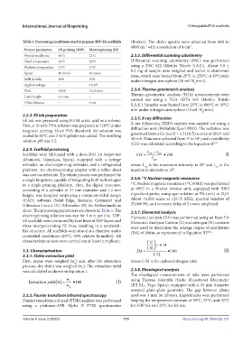
Process parameters 3D printing (3DP) Electrospinning (ES)
Nozzle/needle size 16 G 22 G 2.3.3. Differential scanning calorimetry
Head temperature 32°C 23°C Differential scanning calorimetry (DSC) was performed
Platform temperature 21°C 21°C using a DSC 822 (Mettler Toledo S.A.E.). About 3.0 ±
Speed 10 mm/s 30 mm/s 0.2 mg of sample were weighed and sealed in aluminum
pans, which were heated from 25°C to 250°C at 10°C/min
Infill density 30% 10% under nitrogen atmosphere (10 mL N /min).
Applied voltage – 9.5 kV 2
Flow 155% 12 µL/min 2.3.4. Thermo-gravimetric analysis
Layer height 0.3 mm – Thermo-gravimetric analysis (TGA) measurements were
carried out using a TGA SDTA 851 (Mettler Toledo
Offset distance – 9 cm
S.A.E.). Samples were heated from 25°C to 800°C at 10°C/
min under nitrogen atmosphere (10 mL N /min).
2
2.2.3. ES ink preparation
ES ink was prepared using 0.5 M acetic acid as a solvent. 2.3.5. X-ray diffraction
First, a 10 wt% PVA solution was prepared at 120°C under X-ray diffraction (XRD) analysis was carried out using a
magnetic stirring. Once PVA dissolved, the solution was diffraction unit (PANalyticXpert PRO). The radiation was
cooled to 85°C and 2 wt% gelatin was added. The resulting generated from a Cu-Kα (λ = 1.5418 Å) source at 40 kV and
solution pH was 3.2. 40 mA. Data were collected from 2º to 50º, and crystallinity
(CrI) was calculated according to the Equation II :
[27]
2.2.4. Scaffold processing I − I
Scaffolds were fabricated with a domoBIO 2A bioprinter CrI = 110 am •100 II
(Domotek, Gipuzkoa, Spain), equipped with a syringe I 110
extruder, an electrospinning extruder, and a refrigerated where I is the maximum intensity at 20° and I is the
am
110
platform. An electrospinning adapter with a teflon sheet maximum intensity at 13°.
was used as substrate. The whole process was performed by
13
a single bioprinter, capable of integrating both technologies 2.3.6. C Nuclear magnetic resonance
13
in a single printing platform. First, the digital structure, 13 C Nuclear magnetic resonance ( C NMR) was performed
consisting of a cylinder of 21 mm diameter and 1.2 mm at 90°C in a Bruker Avance unit, equipped with BBO
height, was designed employing a computer-aided design z-gradient probe, using agar solution at 5% (w/v) in D O.
2
(CAD) software (Solid Edge, Siemens, Germany) and About 14,000 scans at 125.75 MHz, spectral window of
Ultimaker Cura 4.13.1 (Ultimaker BV, the Netherlands) as 25,000 Hz, and recovery delay of 2 s were employed.
slicer. The processing parameters are shown in Table 1. The 2.3.7. Elemental analysis
electrospinning solution was run for 1 min per line. 3DP- Elemental analysis (EA) was performed using an Euro EA
ES scaffolds were composed by four lines of 3DP layers and Elemental Analyser. Carbon (C) and nitrogen (N) contents
three interpenetrating ES lines, resulting in a sandwich- were used to determine the average degree of acetylation
like structure. All scaffolds were stored at a chamber under (DA) of chitin, as represented in Equation III :
[28]
controlled conditions (25°C, 50% relative humidity). All
characterization tests were carried out at least in triplicate. C
.
N − 514
2.3. Characterization DA = •100 III
2.3.1. Chitin extraction yield 172.
First, pupae were weighed (w ) and, after the extraction where C/N is the carbon/nitrogen ratio.
p
process, dry chitin was weighed (w ). The extraction yield
c
was calculated as shown in Equation I. 2.3.8. Rheological analysis
The rheological measurements of inks were performed
w using Thermo Scientific Haake Rheostress1 Rheometer
Extraction yield % () = c •100 (I)
w p (IFI S.L., Vigo, Spain), equipped with a 35 mm diameter
serrated plate–plate geometry. The gap between plates
2.3.2. Fourier transform infrared spectroscopy used was 1 mm for all tests. Experiments were performed
Fourier transform infrared (FTIR) analysis was performed keeping the temperature constant at 30°C, 32°C, and 35°C
using a platinum-ATR Alpha II FTIR spectrometer for 3DP ink and 25°C for ES ink.
Volume 9 Issue 3 (2023) 175 https://doi.org/10.18063/ijb.701