
Page 309 - IJB-9-3
P. 309
International Journal of Bioprinting Bioprinted stem cell niche regenerates muscles
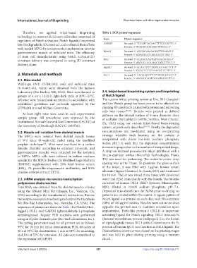
Therefore, we applied inkjet-based bioprinting Table 1. PCR primer sequences
technology to create artificial stem cell niches comprised of Gene Primer sequence
regulators of Notch activation (Notch ligands) bioprinted
into biodegradable 3D construct, and evaluated their effects GAPDH Forward: 5ʹ-TCCATGACAACTTTGGCATTG-3ʹ
with seeded MPCs by intramuscular implantation into the Reverse: 5ʹ-TCACGCCACAGCTTTCCA-3ʹ
gastrocnemius muscle of mdx/scid mice. The efficiency Notch1 Forward: 5ʹ-GCCGCAAGAGGCTTGAGAT-3ʹ
Reverse: 5ʹ-GGAGTCCTGGCATCGTTGG-3ʹ
of stem cell transplantation using Notch activator/3D
construct delivery was compared to using 3D construct Hes1 Forward: 5ʹ-CCAGCCAGTGTCAACACGA-3ʹ
Reverse: 5ʹ-AATGCCGGGAGCTATCTTTCT-3ʹ
delivery alone.
Jagged1 Forward: 5ʹ-ACAGTTGTTATGGGTGGCTCT-3ʹ
Reverse: 5ʹ-CGGCTCCTCTCACGTTCTTTC-3ʹ
2. Materials and methods DLL1 Forward: 5ʹ-CAGGACCTTCTTTCGCGTATG-3ʹ
2.1. Mice model Reverse: 5ʹ-AAGGGGAATCGGATGGGGTT-3ʹ
Wild-type (WT; C57BL/10J), mdx and mdx/scid mice
(6-month-old, males) were obtained from the Jackson
Laboratory (Bar Harbor, ME, USA). Mice were housed in 2.4. Inkjet-based bioprinting system and bioprinting
groups of 4 on a 12:12-h light-dark cycle at 20°C–23°C. of Notch ligand
All mice were housed and maintained in accordance with The custom inkjet printing system at Drs. Phil Campbell
established guidelines and protocols approved by the and Lee Weiss’s group has been proven to be efficient for
UTHealth Animal Welfare Committee. creating 3D constructs printed with protein and delivering
cells into tissues [40-43] . Bioinks were printed as defined
At least eight mice were used in each experimental patterns on the dermal surface of 4-mm diameter discs
sample group. All procedures were approved by the of acellular DermaMatrix (ADM; Synthes, West Chester,
Institutional Animal Care and Use Committee (IACUC) at PA, USA) using our custom inkjet-based biopatterning
the University of Pittsburgh (IACUC-1109718). system, as previously described [40-43] . Briefly, the deposited
2.2. Muscle cell isolation from skeletal muscle concentrations are modulated using an overprinting
The MPCs were isolated from skeletal muscle tissues strategy whereby each location on the pattern is
of WT mice (8-week-old, male) using the modified overprinted with dilute bio-inks (sodium phosphate
preplate technique . Mice were sacrificed in a carbon buffer, pH 7.4) such that the deposited concentrations
[29]
dioxide chamber according to standard protocols, and increase in proportion to the number of overprinted drops.
gastrocnemius muscles were collected for the isolation A drop-on-demand piezoelectric inkjet printhead with a
of MPCs. MPCs cells were cultured in culture medium 30-μm-diameter orifice (MicroFab Technologies, Plano,
specific for the MPCs (Dulbecco’s Modified Eagle Medium TX) was used for patterning. The center-to-center drop
[DMEM] supplemented with 20% fetal bovine serum spacing was set to 75 μm. To passivate the glass surface
[FBS], 1% penicillin-streptomycin antibiotics, and 0.5% of the inkjet, it was filled with 1 μg/mL bovine serum
chicken embryo extract [CEE]). albumin (Sigma Chemical, St. Louis, MO) and incubated
for 10 min. The jet was rinsed three times with deionized
2.3. mRNA analysis via reverse-transcription water and filled immediately with the bioink. The bioinks
polymerase chain reaction consisted of human DLL1 (R&D Systems, Minneapolis,
Total RNA was obtained from the skeletal muscles of mice MN), diluted in 10 mM sodium phosphate, pH 7.4.
using the RNeasy Mini Kit (Qiagen, Inc., Valencia, CA, Deposited inks absorb into the ADM prior to drying, so
USA) according to the manufacturer’s instructions. Reverse patterns are created within the matrix. A square pattern of
transcription was performed using an iScript cDNA Synthesis Notch ligand was printed on each disc, with 50 overprints
Kit (Bio-Rad Laboratories, Inc., Hercules, CA, USA). The (OPs) of 100 μg/ml bioinks. Notches were cut in the discs
sequences of primers are shown in Table 1 for Notch1, Hes1, opposite the printed area to maintain orientation upon
Jagged1, DLL1, and GAPDH (glyceraldehyde 3-phosphate implantation. Delta-like protein-1 (DLL1) is important
dehydrogenase). Regular PCR reactions were performed activating ligand for Notch signaling. DLL1 (mouse):Fc
using an iCycler thermal cycler (Bio-Rad Laboratories, Inc.). (human) recombinant protein (Adipogen) (i.e., the fusion
The cycling parameters used for all primers are as follows: of signal peptide mouse DLL1 at the C-terminus to the Fc
95°C for 10 min for initial denaturation; PCR, 40 cycles of portion of human IgG1) can function as DLL1 ligand. The
30 s at 95°C for denaturation, 1 min at 54°C for annealing, DermaMatrix construct was placed on the printing stages
and 30 s at 72°C for extension. All data were normalized to and was held in place during printing using a vacuum
the expression of GAPDH. chuck.
Volume 9 Issue 3 (2023) 301 https://doi.org/10.18063/ijb.711