
Page 311 - IJB-9-3
P. 311
International Journal of Bioprinting Bioprinted stem cell niche regenerates muscles
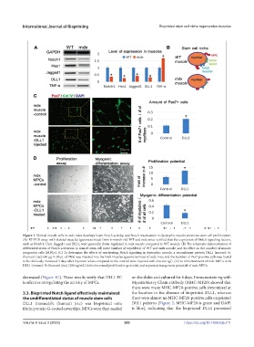
Figure 1. Skeletal muscle cells in mdx mice develops lower Notch activity, and Notch reactivation in dystrophic muscle promotes stem cell proliferation.
(A) RT-PCR assay with skeletal muscles (gastrocnemius) from 6-month-old WT and mdx mice verified that the expression of Notch signaling factors,
such as Notch1, Hes1, Jagged1 and DLL1, were generally down-regulated in mdx muscle compared to WT muscle. (B) The schematic demonstration of
differential status of Notch activation in muscle stem cell niche (surface of myofibers) of WT and mdx muscle, and its effect on the number of muscle
progenitor cells (MPCs). (C) To determine the effects of reactivating Notch signaling in dystrophic muscle, a recombinant protein DLL1 (mouse): Fc
(human) (rec) (40 µg in 20 µL of PBS) was injected into the limb muscles (gastrocnemius) of mdx mice and the number of Pax7-positive cells was found
to be obviously increased 3 days after injection when compared to the control mice injected with inactive IgG. (D) In vitro treatment of mdx MPCs with
DLL1 (mouse): Fc (human) (rec) (200 ng/mL) led to increased proliferation potential and repressed myogenesis potential of mdx MPCs.
decreased (Figure 1C). These results verify that DLL1-FC on the slides and cultured for 4 days. Immunostaining with
is effective at regulating the activity of MPCs. Myosin Heavy Chain antibody (MHC-MF20) showed that
there were many MHC-MF20-positive cells (myotubes) at
3.3. Bioprinted Notch ligand effectively maintained the location in the absence of bioprinted DLL1, whereas
the undifferentiated status of muscle stem cells there were almost no MHC-MF20-positive cells on printed
DLL1 (mouse):Fc (human) (rec) was bioprinted onto DLL1 patterns (Figure 2, MHC-MF20 is green and DAPI
fibrin/protein G-coated coverslips. MPCs were then seeded is blue), indicating that the bioprinted DLL1 prevented
Volume 9 Issue 3 (2023) 303 https://doi.org/10.18063/ijb.711