
Page 92 - IJB-9-4
P. 92
International Journal of Bioprinting Laser transfer for CTC isolation
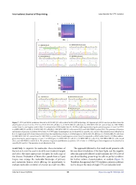
Figure 7. TP53 and BRAF mutations detected in MDA-MB-231 cells isolated by BA-LIFT technology. (A) Agarose gel of PCR reaction products from the
amplification of exon 9 of the TP53 in a MDA-MB-231 cell (lane 1), 25 MDA-MB-231 cells (lane 2), 1000 MDA-MB-231 control (lane 3), 1000 PBMCs
control (lane 4), and negative control (lane 5) compared to a DNA ladder (lane L). (B) Electropherogram showing the partial sequence of exon 9 of TP53
in a MDA-MB-231 cell (B.1), 25 MDA-MB-231 cells (B.2), 1000 MDA-MB-231 cells control (B.3), and 1000 PBMCs control (B.4). The presence of thymine
and absence of guanine at position 839 of exon 9 of TP53 gene (homozygous) can be observed in cases B.1, B.2, and B.3. The mutation is not observed in
B.4. (C) Agarose gel of PCR reaction products from the amplification of exon 11 of BRAF gene in a MDA-MB-231 cell (lane 1), 25 MDA-MB-231 cells (lane
2), 1000 MDA-MB-231 control (lane 3), 1000 PBMCs control (lane 4), and negative control (lane 5) compared to a DNA ladder (lane L). (D) Electrophero-
gram showing the partial sequence of exon 11 of BRAF gene in a MDA-MB-231 cell (D.1), 25 MDA-MB-231 cells (D.2), 1000 MDA-MB-231 cells control
(B.3), and 1000 PBMCs control (B.4). The presence of guanine and thymine at position 1391 of exon 11 of BRAF gene (heterozygous) can be observed in
cases B.1, B.2 and B.3. The mutation is not observed in B.4.
would help to improve the molecular characterization of The approach followed in this work would preserve cells
the tumor, it could be used to identify new treatment targets for any direct irradiation of the laser light, and the negative
and select the most appropriate therapies for each stage of selection approach followed would maintain the CTCs free of
the disease. Evaluation of biomarker panels from a liquid any direct labeling, preserving the cells in perfect condition
biopsy may enlarge the molecular landscape of primary for further culture, characterization, or analysis (Figure 9).
and metastatic lesions while offering the opportunity to Therefore, this approach for CTCs isolation presents a relevant
evaluate molecular evolution of a tumor as a real-time film. tool to deepen the study of single CTCs at molecular level.
Volume 9 Issue 4 (2023) 84 https://doi.org/10.18063/ijb.720