
Page 218 - IJB-9-5
P. 218
International Journal of Bioprinting Hydrogels for 3D bioprinting
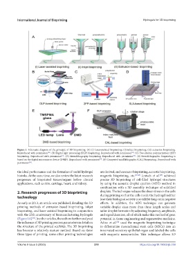
Figure 1. Schematic diagram of the principle of 3D bioprinting. (A) (i) Laser-assisted bioprinting; (ii) inkjet bioprinting; (iii) extrusion bioprinting.
Reproduced with permission . (B) Digital light processing (DLP) bioprinting. Reproduced with permission . (C) Two-photon polymerization (2PP)
[34]
[26]
bioprinting. Reproduced with permission . (D) Stereolithography bioprinting. Reproduced with permission . (E) Stereolithographic bioprinting is
[32]
[33]
based on the digital micromirror device (DMD). Reproduced with permission . (F) Computed axial lithography (CAL) bioprinting. Reproduced with
[35]
permission .
[38]
the ideal performance and the formulas of useful hydrogel are derived, such as nano-bioprinting, acoustic bioprinting,
bioinks. At the same time, we also review the latest research magnetic bioprinting, etc. [27,28] Jentsch et al. achieved
[29]
progresses of bioprinted tissues/organs before clinical precise 3D bioprinting of cell-filled hydrogel structures
applications, such as skin, cartilage, heart, and kidney. by using the acoustic droplet ejection (ADE) method in
combination with a 3D assembly technique of solidified
2. Research progresses of 3D bioprinting droplets. The technique reduces the shear stress on the cells
technology during printing so that the cells inside the hydrogel neither
lose their biological activity nor exhibit long-term negative
As early as 2013, an article was published, detailing the 3D effects. In addition, the ADE technique can generate
printing methods of extrusion-based bioprinting, inkjet variable droplet sizes more than three length scales and
bioprinting, and laser-assisted bioprinting in conjunction tailor droplet formation by adjusting frequency, amplitude,
with the 25th anniversary of biomanufacturing hydrogels and signal duration, all of which make this method of great
(Figure 1A) . In other articles, the authors further analyzed potential in tissue engineering and regenerative medicine.
[26]
the influence of 3D printing process parameters in detail on Adine et al. used the magnetic bioprinting technique
[30]
the structure of the printed scaffolds. The 3D bioprinting to differentiate mesenchymal stem cells (MSCs) into an
has become a relatively mature method. Based on these innervated secretory epithelial organ and labeled the cells
three types of printing, some other printing technologies with magnetic nanoparticles. This technique allows 3D
Volume 9 Issue 5 (2023) 210 https://doi.org/10.18063/ijb.759