Page 332 - IJB-9-5
P. 332
International Journal of Bioprinting Review of 4D-printed smart medical implants
that underwent 4D cyclic mechanical loading proved
References to have an influence on fibroblast-to-myofibroblast
transdifferentiation
. Moreover, Douillet et al. presented
[120]
a novel model via laser-assisted bioprinting (LAB) to make
[122]
[124]
[109]
[121]
[126]
[90]
a replication of fibroblast dynamic remodeling in vitro [121] .
This method can be utilized to understand better cellular
behaviors and interactions with the extracellular matrix
Possible applications Cartilage-like tissue Cartilage-like tissue Soft tissue engineering Mimicking microvessels Scaffolds for tissue engineering Culture substrates (ECM). Except for these studies that affect cells through
the regulation of internal networks, some researchers use
cell-laden 4D bioprinting to form cartilage-like tissue
[90,122]
, which are quite meaningful to
and vascularized tissue
[123]
tissue/organ regeneration applications.
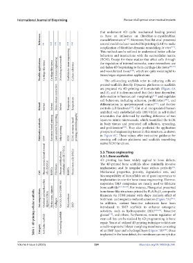
The cell-seeding scaffolds refer to culturing cells on
Stress-release contraction printed scaffolds directly. Dynamic platforms or scaffolds
are prepared via 4D printing of biomaterials (Figure 6A
and B), and it is demonstrated that their time-dependent
and regulates
deformation influences cell morphology
[124]
, and
cell behaviors, including adhesion, proliferation
[125]
Stimuli Solvent Solvent Solvent Temperature Temperature differentiation in spatiotemporal control [126] , and further
[127]
controls cell functions
. Cui et al. encapsulated human
umbilical vein endothelial cells (HUVECs) in self-folded
microtubes that deformed by swelling difference of two
layers to mimic microvessels, which resembled the ECM
Fabrication methods Extrusion-based printing Laser-assisted bioprinting Inkjet printing FDM, extrusion-based printing, stereolithography in body tissues and promoted cell adhesion, spreading,
and proliferation
. They also predicted the application
[109]
prospects of engineering tissues in this structure, as shown
in Figure 6C. These values offer instructive guidance for
FDM
DIW
creating cell culture platforms and scaffolds resembling
native ECM functions.
Human mesenchymal stromal Human umbilical vein endothelial cells (HUVECs) Neural stem cells (NSCs) 5.3. Tissue engineering
5.3.1. Bone scaffolds
4D printing has been widely applied to bone defects.
The 4D-printed bone scaffolds allow minimally invasive
[23]
Mechanical properties, porosity, degradation rate, and
Cell cells (hMSCs) hMSCs Fibroblasts hMSCs implantation and fit irregular bone defects perfectly .
biocompatibility of bioscaffolds are of great importance to
implantation in vivo for bone tissue engineering. Thermo-
Oxidized and methacrylate
responsive SMP composites are mostly used to fabricate
bone scaffolds
. For instance, Zhang et al. presented
[116,128,129]
Table 1. 4D printing cellular scaffolds. Ink compositions Cell-laden scaffolds alginate (OMA) Hyaluronan, alginate Collagen Gel-MA, Gel-COOH-MA Cell-seeding scaffolds Polyurethane SMP filaments via FDM printer with shape memory effect of
bone tissue-like structures printed by PLA/Fe O composite
3
4
.
both heat- and magnetic-induced actuation (Figure 7A)
[116]
In addition, various bioactive substances have been
introduced to SMP scaffolds to enhance osteogenic
, bioactive
activities, such as hydroxyapatite (HA)
[130,131]
glasses
, and others. Furthermore, remote regulation of
[132]
stem cell fate can be realized by 4D programming in bone
repair. You et al. utilized 4D printing technique to fabricate
Category
a multi-responsive bilayer morphing membrane consisting
implanted in the bone defect, the membrane can morph due
Volume 9 Issue 5 (2023) 324 of an SMP layer and a hydrogel layer (Figure 7B) [108] . Once
https://doi.org/10.18063/ijb.764

