
Page 351 - IJB-9-5
P. 351
International Journal of Bioprinting 3D printing of tough and self-healing hydrogels
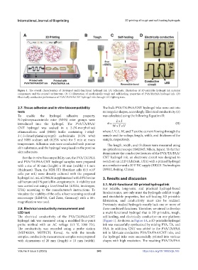
Figure 1. The overall characteristics of developed multi-functional hydrogel ink. (A) Schematic illustration of 3D-printable hydrogel ink material
components and the printed architecture. (B, C) Illustration of mechanically tough and self-healing properties of PVA/TA/PAA hydrogel ink. (D)
Electrically conductive performance of PVA/TA/PAA/CNT hydrogel inks through LED lighting tests.
2.7. Tissue adhesion and in vitro biocompatibility The bulk PVA/TA/PAA/CNT hydrogel inks were cut into
tests rectangular shapes, accordingly. Electrical conductivity (σ)
To enable the hydrogel adhesive property, was calculated using the following Equation II:
N-hydroxysuccinimide ester (NHS) ester groups were LI
introduced into the hydrogel. The PVA/TA/PAA/ WT (II)
V
CNT hydrogel was soaked in a 2-(N-morpholino)
ethanesulfonic acid (MES) buffer containing 1-ethyl- where I, V, L, W, and T are the current flowing through the
3-(-3-dimethylaminopropyl) carboiimide (0.5% w/w) sample and the voltage, length, width, and thickness of the
and NHS sodium salt (0.25% w/w) for 5 min at room sample, respectively.
temperature. Adhesion tests were conducted with porcine The length, width, and thickness were measured using
skin substrates, and the hydrogel was placed on the porcine an optical microscope (SMZ645; Nikon, Japan). To further
skin substrates. demonstrate the conductive features of the PVA/TA/PAA/
For the in vitro biocompatibility test, the PVA/TA/PAA CNT hydrogel ink, an electronic circuit was designed to
and PVA/TA/PAA/CNT hydrogel samples were prepared switch on an LED (Adafruit, USA) with a printed hydrogel
with a size of 10 mm (height) × 10 mm (width) × 1 mm as a conductor and a 30 V DC supply (RIGOL Technologies
(thickness). Then, the NIH-3T3 fibroblast cells (0.5 ×10 DP832, Beijing, China).
5
cells per mL) were directly cultured with the prepared
hydrogel in 1 mL of DMEM supplemented with 10% bovine 3. Results and discussion
calf serum and 1% penicillin–streptomycin. A viability test
was carried out using a Live/Dead kit (L3224, Invitrogen, 3.1. Multi-functional 3D-printed hydrogel ink
USA) according to the manufacturer’s instructions. To For reliable, long-term, and practical hydrogel-based
visualize the viability of the cells, a laser scanning confocal bioelectronics, not only must the hydrogels exhibit tough
microscope (LSM700, Carl Zeiss, Germany) with a 10× and stretchable properties, but self-healing 3D printable
magnification was used. fabrication, and conductivity must also be realized.
Previously studied hydrogels mostly lack one or more of
2.8. Electrical conductivity measurement and these combined functions. Therefore, we aimed to develop
LED test a multi-functional hydrogel that is 3D printable, tough,
The electrical conductivity of the PVA/TA/PAA/CNT self-healing, and electrically conductive on one platform
hydrogel ink was measured using a modified four-point (Figure 1). As shown in Figure 1A, a 3D printable hydrogel
probe method with an AC voltage (±1 V, 1000 Hz). ink was successfully synthesized by mixing PVA, TA, and
The conductivity was recorded using a probe station PAA. In addition, CNT was added to the PVA/TA/PAA
(MST4000A, MSTECH, Korea). As with the tensile ink to fabricate conductive PVA/TA/PAA/CNT inks, and
samples, conductivity measurement samples were prepared the hydrogel inks were successfully printed into various
with dimensions of 20 mm (length) × 10 mm (width). shapes with high resolution. The resulting PVA/TA/PAA
Volume 9 Issue 5 (2023) 343 https://doi.org/10.18063/ijb.765