
Page 356 - IJB-9-6
P. 356
International Journal of Bioprinting Surface modification of PCL scaffolds
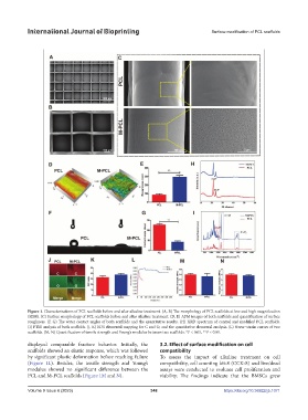
Figure 1. Characterizations of PCL scaffolds before and after alkaline treatment. (A, B) The morphology of PCL scaffolds at low and high magnification
(SEM). (C) Surface morphology of PCL scaffolds before and after alkaline treatment. (D, E) AFM images of both scaffolds and quantification of surface
roughness. (F, G) The water contact angles of both scaffolds and the quantitative results. (H) XRD spectrum of control and modified PCL scaffolds.
(I) FTIR analysis of both scaffolds. (J, K) EDS elemental mapping for C and O, and the quantitative elemental analysis. (L) Stress−strain curves of two
scaffolds. (M, N) Quantification of tensile strength and Young’s modulus between two scaffolds. *P < 0.05, **P < 0.01.
displayed comparable fracture behavior. Initially, the 3.2. Effect of surface modification on cell
scaffolds showed an elastic response, which was followed compatibility
by significant plastic deformation before reaching failure To assess the impact of alkaline treatment on cell
(Figure 1L). Besides, the tensile strength and Young’s compatibility, cell counting kit-8 (CCK-8) and live/dead
modulus showed no significant difference between the assays were conducted to evaluate cell proliferation and
PCL and M-PCL scaffolds (Figure 1M and N). viability. The findings indicate that the BMSCs grew
Volume 9 Issue 6 (2023) 348 https://doi.org/10.36922/ijb.1071