
Page 269 - v11i4
P. 269
International Journal of Bioprinting 3D cell culture model for neural cell analysis
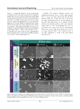
(Figure 1). Sequential filtration using polycarbonate Similarly, ZTA particles exhibited granular and
membrane filters (8, 0.8, and 0.1 μm pore sizes) categorized polygonal morphology (Figure 1), with a mean diameter
particles into >8, 0.8–8, and 0.1–0.8 μm size ranges. SEM of 3.7 ± 3.5 μm and a size range of 0.45–20.3 μm. Most
analysis revealed that PEEK-OPTIMA™ particles exhibited particles (99%) were <8 μm, with 73% in the 0.8–8.0
a granular or irregular morphology across all size ranges μm range. Additionally, 99.5% of the total particle area
(Figure 1), with a mean diameter of 7.6 ± 3.9 μm and a size was within this range (Figure 2c). In contrast, CoCrMo
range of 0.54–83.2 μm. While 95% of particles were <8 μm, particles were primarily globular, with some polygonal
43% were in the submicron range. Larger particles (>8 μm)
accounted for ~50% of the total particle area (Figure 2a). shapes (Figure 1). Their mean diameter was 5.9 ± 1.2
®
Ceridust 3615 particles displayed a granular and polygonal μm, within a size range of 2.1–11.6 μm. Most particles
morphology (Figure 1), with a mean diameter of 6.4 ± (88%) were <8 μm, with 88.5% falling in the 0.8–8.0
2.7 μm and a size range of 0.49–22.1 μm. Approximately, μm range. However, while 11.5% of particles were >8
92.3% of particles were <8 μm, with a notable proportion μm, they contributed to 56.2% of the total particle
(1%) in the 0.1–0.8 μm range (Figure 2b). area (Figure 2b).
Figure 1. SEM images of model particles, including aggregates and microsized particles, collected using 8, 0.8, and 0.1 μm filters. A consistent scale bar
of 10 μm was applied across all SEM images (each filter size) for uniform comparison. Abbreviations: CoCrMo: Cobalt–chromium–molybdenum; PEEK:
Polyetheretherketone; SEM: Scanning electron microscopy; ZTA: Zirconia-toughened alumina.
Volume 11 Issue 4 (2025) 261 doi: 10.36922/IJB025180174