
Page 272 - v11i4
P. 272
International Journal of Bioprinting 3D cell culture model for neural cell analysis
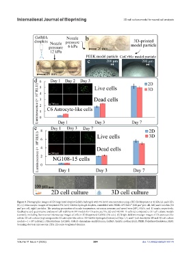
Figure 3. Photographic images of 3D-bioprinted droplet GelMA hydrogels with 5% (w/v) concentration using a BIO X6 bioprinter at 12 kPa (a) and 6 kPa
(b). (c) Microscopic images of bioprinted 5% (w/v) GelMA hydrogel droplets, embedded with PEEK-OPTIMA™ (100 μm³ per cell, left) and CoCrMo (50
μm³ per cell, right) particles. The printing parameters of nozzle temperature, extrusion pressure, and speed were 24°C, 6 kPa, and 11 mm/s, respectively.
Qualitative and quantitative analyses of cell viability in 3D models for C6 astrocyte-like (d) and NG108-15 cells (e) compared to 2D cell culture models
(control), including fluorescence microscopy images of cells in 3D-bioprinted GelMA (5% w/v). (f) Bright-field microscope images of C6 astrocyte-like
cells in 2D cell culture (top) compared to C6 astrocyte-like cells in 3D GelMA hydrogels (bottom) at Days 1, 3, and 7 (cell density for 2D and 3D cell culture
4
models = 1 × 10 cells/mL). Abbreviations: CoCrMo: Cobalt–chromium–molybdenum; GelMA: Gelatin methacryloyl; PEEK: Polyetheretherketone; SEM:
Scanning electron microscopy; ZTA: Zirconia-toughened alumina.
Volume 11 Issue 4 (2025) 264 doi: 10.36922/IJB025180174