
Page 18 - ITPS-5-2
P. 18
12 INNOSC Theranostics and Pharmacological Sciences, 2022, Vol. 5, No. 2 Yanik et al.
Rapid diagnosis and treatment of bacterial half of the cornea. The anterior chamber, pupil, and
keratitis are essential to limit stromal scarring and lens looked normal. The fundus was not enlightened.
minimize potential vision loss. Thus, treatment Left corneal scraping material and cultures
should be started empirically early [6]. This were taken. Gram stains of the samples from the
pathogen is mostly seen in adults due to contact conjunctiva and cornea were performed. Abundant
lens usage [7]. However, the most important risk leukocytes and Gram-negative bacillus were seen
is hospitalization for infants. In the literature, at the microscopic examination on Gram stains.
P. aeruginosa keratitis in infants is frequently All samples were inoculated with 5% sheep
hospital-acquired. Cases of non-hospital-acquired blood, eosin methylene blue (EMB) agar, and
P. aeruginosa keratitis in infants are especially rare. chocolate agar containing Poly ViteX for aerobic
bacterial cultures and were incubated at 37°C for
2. Case presentation 24–48 h. Samples of EMB and 5% sheep blood
A 7-month-old female infant who had no known agar cultures from the left conjunctiva and left
chronic disease was brought in with the complaint cornea produced typical P. aeruginosa (Figure 2).
of watering and redness on the left eye. One month This pathogen was found to be susceptible to
prior, the patient swam in a pool on a farm, and then amikacin, ceftazidime, levofloxacin, ciprofloxacin,
the complaints started. At that time, nasolacrimal piperacillin, and tazobactam. Gentamicin eye drop
th
duct obstruction was considered by a physician at was added to the treatment on the 4 day of the
the ophthalmologic examination, and the physician treatment.
prescribed some eye drops and suggested waiting The produced P. aeruginosa was detected
1 year. The patient has used netilmicin and using API 10 S Gram-negative identification kit
dexamethasone drops during this time. When the (Biomerieux, ABD). Antibiotic susceptibility tests
patient came to our clinic again, her complaints were performed with disc diffusion (Figure 3).
did not decrease. According to ophthalmologic Clinical and Laboratory Standards Institute criteria
examination results, the patient was diagnosed with
keratitis. Macroscopically, the right eye appeared
normal, and the anterior and posterior chambers
of the right eye were also normal. The left eye
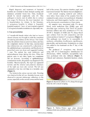
conjunctiva was less red than in a keratitis case
(Figure 1).
The lesion in the cornea was not wide. Watering
was observed in the left eye. Keratitis focuses were
observed to prevent retinoscopy reflections in the
cornea, especially on the central cornea and the lower
Figure 2. Bacterial culture on 5% sheep blood agar
from the left corneal scraping.
Figure 3. Muller–Hinton agar plate showed
Figure 1. Pre-treatment corneal appearance. antimicrobial susceptibility profile of the pathogen.
©2022 AccScience Publishing