
Page 12 - ITPS-7-1
P. 12
INNOSC Theranostics and
Pharmacological Sciences Liquid biopsy and digital PCR in cancer
Blood or other bodily fluids can be used in the early This property even makes it possible to verify the
detection of information about a tumor that perhaps cannot presence of metastasis in the central nervous system in
yet be detected by imaging procedures. The high sensitivity cases of patients duly treated with first-line ITKs, where
of molecular detection methodologies allows LB to become the original tumor is inactive, and is no longer releasing
the ideal tool for monitoring therapeutic responses. Since genetic material into the plasma, but as a consequence
the circulating genetic material can be found as free form in of the pharmacological pressure, sensitive mutations are
plasma, cerebrospinal fluid, urine, pleural fluid, and ascites, still present in circulating DNA in the cerebrospinal fluid.
or as RNA adhered to (protected by) the platelet membranes. Under this circumstance, the originally “sensitive” tumor
Circulating genetic material may also be quantifiable. Thus, managed to generate systemic metastases in different
the “molecular charge” or number of copies of given genes organs that can be abrogated by specific therapy, except
detected by multiplex droplet digital polymerase chain those in the central nervous system since the access of the
reaction (ddPCR) procedure is proportional to the tumor drugs is limited by the blood-brain barrier due to the high
[38]
mass that is producing it . In this way, it is possible to verify expression of the ABC-t of MDR. All positive results in the
the drop in the number of copies of sensitive mutations, but detection of one or several of these somatic mutations can
at the same time, detect resistant mutations after a period identify and characterize a tumor constitute quantifiable
of treatment with first-line ITKs. Clearly, this simultaneous markers, which can be used to detect minimal residual
information gives us an idea of the degree of efficacy of the disease. Other important information is the presence
first treatment, and documents the presence of a tumor of hypermethylated DNA fragments in CpG islands of
relapse at the expense of a change in the pharmacogenetic promoter regions of tumor suppressor genes, such as
identity of the emerging clone of said tumor. These SEPT9 (colon cancer) or SHOX2 (lung cancer). This type
“mutational changes” produced in the original clone, largely of epigenetic silencing, which inhibits tumor apoptosis,
to be expected after the pharmacological pressure is exerted, serves as an excellent marker of tumor lineage (Figure 5).
provide new therapeutic targets that can be acted upon with
other second- or third-generation drugs in some cases. Thus, 5. ddPCR applied to LB
targeted therapy could be started long before the cancer is At present, the latest ddPCR amplification techniques
clinically evident or detected by imaging. In general, when increases the sensitivity of detection, obtaining positive
the images appear, it is because there is an important tumor results even when the copy load of each of the mutations
mass and often consistent with stages of dissemination. sought is extremely low. Under this circumstance, the
tumor mass will be <10 cells, and the imaging studies will
4
Figure 4. Every tumor usually presents a heterogeneous cell mass, and its
genetic information will be present in the circulation. Circulating tumor
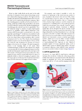
Figure 3. Liquid biopsy, implemented with droplet digital polymerase cells, exosomes and different DNA fragments can be detected, quantified
chain reaction, manifests the characteristics of an ideal biomarker. With and identified according to the specific information they carry (i.e.,
high specificity and sensitivity, it can detect mutations of clonal lineage as immuophenotype and somatic mutations). The same information can be
well as sensitivity and resistance to treatment. The biomarker should be detected in virtually all bodily fluids. Furthermore, larger DNA fragments
quantifiable, giving an idea of the magnitude of the tumor size, as well as without specific genetic information are derived from cell turnover in
serving as an indicator of minimal residual disease. normal tissues.
Volume 7 Issue 1 (2024) 6 https://doi.org/10.36922/itps.1227