
Page 10 - JCBP-1-1
P. 10
Journal of Clinical and
Basic Psychosomatics Brain MRI alterations in MDD
monoamines originate from the median suture nucleus,
locus coeruleus, and substantianigra and ventral tegmental
area, respectively [3,4] .
However, diagnostic biomarkers of MDD are still
lacking. Current first-line treatments show moderate
efficacy, with clinical response at around 50 – 60%
and remission at approximately 20 – 30% . Clinical
[9]
studies have found that the response rate to initial
antidepressants is approximately 50% . Thus, describing
[10]
the neurobiological basis of MDD is expected to support
the development of more effective diagnostic methods
and treatments.
Magnetic resonance imaging (MRI), an intuitive and
non-invasive tool for visualizing the structure and function
of the brain, can be particularly helpful in understanding
psychiatric disorders and the relevant treatment responses. Figure 1. Science citation index expanded publications on magnetic
Although the pathophysiology of MDD has been resonance imaging in depression and antidepressant treatment.
intensively studied using MRI, clinical imaging markers
for diagnosis and predictors of treatment outcome have not structural MRI techniques used to assess regional gray
been identified. This review describes the structural and matter volumes, and diffusion tensor imaging (DTI) used
functional brain changes after antidepressant treatment to reflect microstructural changes in the white matter,
in MDD using MRI and identifies possible neuroimaging allowing visualization of structural changes and differences
biomarkers in MDD. in brain tissue composition.
2. Literature search Some DTI studies have shown reduced fractional
anisotropy (FA) values in the radial corona, corpus
This narrative review focuses on the studies of callosum, superior longitudinal fasciculus, hooked
pharmacological treatment-associated brain structural fasciculus, internal capsule, cingulate gyrus, amygdala,
and functional alterations in MDD that were identified and orbitofrontal lobe in patients with MDD, and
by searching the PubMed and Web of Science further studies have shown that lower FA values in the
databases from 2000 to September 2022. Keywords left internal capsule are associated with higher depressive
used in combination included “MRI,” “structural MRI,” severity [11,12] . A study on the structural network of
“functional MRI (fMRI),” “MDD,” “depression,” and cerebral white matter using 7.0T MRI identified
“antidepressants.” The identified articles were then significantly decreased hippocampal connectivity in
cross-referenced. Structural and fMRI studies were patients with MDD compared to healthy controls .
[13]
included if they (i) were written in English and (ii) had Longitudinal studies revealed a significant increase in FA
human study approval information. Case reports were values in the left superior longitudinal fasciculus, inferior
excluded from the study. Studies that included patients longitudinal fasciculus, cingulate gyrus, and bilateral
with remitted depression rather than ongoing MDD, uncinate fasciculus in patients with MDD after treatment,
tested experimental pharmacological interventions, and the researchers have found a positive correlation
investigated treatment-resistant patients, or were not between FA values before treatment and the efficacy of
recruited in the expanded science index were excluded antidepressants [14-16] .
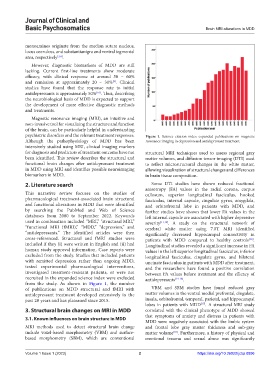
from the study. As shown in Figure 1, the number
of publications on MDD structural and fMRI with VBM and SBM studies have found reduced gray
antidepressant treatment developed extensively in the matter volumes in the ventral medial prefrontal, cingulate,
past 20 years and has plateaued since 2015. insula, orbitofrontal, temporal, parietal, and hippocampal
lobes in patients with MDD . A structural MRI study
[17]
3. Structural brain changes on MRI in MDD correlated with the clinical phenotype of MDD showed
that symptoms of anxiety and distress in patients with
3.1. Known influences on brain structure in MDD
MDD were negatively associated with the limbic system
MRI methods used to detect structural brain change and frontal lobe gray matter thickness and sub-gray
include voxel-based morphometry (VBM) and surface- matter volume . Furthermore, a history of physical and
[18]
based morphometry (SBM), which are conventional emotional trauma and sexual abuse was significantly
Volume 1 Issue 1 (2023) 2 https://doi.org/10.36922/jcbp.0896