
Page 96 - JCBP-3-1
P. 96
Journal of Clinical and
Basic Psychosomatics Functional neurological movement disorders
with a distinctive set of predisposing factors at play. It has
3
been reported that patients with psychogenic movement
disorder reported higher rates of childhood trauma,
specifically greater emotional abuse and physical neglect,
greater fear associated with traumatic events, and a greater
number of traumatic episodes compared with healthy
volunteers. However, ongoing physical or psychological
2
traumas, infections, and medical disorders may precipitate
functional disorders. General cognitive functions are
4,5
found to be normal in patients with functional movement
disturbances in contrast to psychogenic non-epileptic
6,7
seizures. About 15% of patients referred to outpatient
neurological clinics are suffering from neurological
functional disorders. These patients are often discovered
8
to present additional symptoms from other organ systems
such as the gastrointestinal system, heart, and lungs.
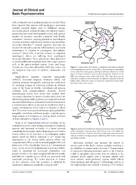
Females are more often affected by FMD than males, and Figure 1. Coronal view of the brain. A voluntary movement is initiated
the symptoms may occur in children, adolescents, and in the supplementary motor cortex (SMC). The impulses are transmitted
adults. to the cerebellum, basal ganglia, and nigral substance (blue lines), which
adjust the body movement, and returned through the thalamus to the
Single-photon emission computed tomography SMC and primary motor cortex (red lines). This long circle may be
(SPECT), functional magnetic resonance (fMR), and influenced possibly at several locations by inhibitory impulses from the
limbic system. Moreover, there is a reciprocal connection between SMC
positron emission tomography scanning are useful tools and the primary motor cortex.
for studying changes in activation patterns in different
parts of the brain in healthy individuals and persons
suffering from neuropsychiatric disorders. Several
neuroimaging studies have shown that multiple brain
structures, important for motor activation and control, are
affected in patients suffering from neurological functional
movement disturbances. Increased activation of neurons in
a restricted area reflects an increase in metabolism, that is,
oxygen consumption, which leads to an increase in blood
flow. The motor cortex, prefrontal areas, right parietal lobe,
amygdala, cerebellum and basal ganglia, cingulate gyrus,
hippocampus, and thalamus, are, among others, activated
in abnormal patterns (Figures 1 and 2).
Stone et al. demonstrated reduced activation of the
9
contralateral motor cortex during movement of weakened
muscles and increased activation of a broad network Figure 2. Sagittal view of the brain. Increased activity in parts of the limbic
comprising basal ganglia, insula, lingual gyri, and inferior system (amygdala, cingulate gyrus, orbitofrontal cortex, and insula) may
frontal cortex. In an overview of neuroimaging results, disturb normal motor function. Memory of unpleasant events may lead to
10
mainly based on SPECT, Girouard et al. found that reduced activity in the hippocampus, indicating a link between memory
and limbic overload.
hypoactivity at the frontal (46%), parietal (38%), and
temporal parts (29%), as well as in basal ganglia (29%) and Emotional overload may increase the activity in
brain stem (17%). Specifically, Voon et al. demonstrated several parts of the limbic system, probably resulting in
11
lower activity in the left supplementary motor area (SMA), an inhibition of normal motor function. 13,14 Improvement
which is important for movement preparation, and of symptoms following treatment seems to be linked to
greater activity in the right amygdala left anterior insula, a shift in amygdala connectivity from its relation to the
and bilateral cingulate gyrus, which are associated with posterior motor regions onto a dynamic relation with
emotional processing. The motor intension is normal more anterior motor/prefrontal regions, which reflects
15
on the conscious level, but execution is inhibited by better planning and self-agency. Interestingly, activity in
modulatory influences outside the conscious will. 12 the hippocampus was reduced, indicating a link between
Volume 3 Issue 1 (2025) 90 doi: 10.36922/jcbp.4369