
Page 8 - JCTR-10-1
P. 8
4 Lv et al. | Journal of Clinical and Translational Research 2024; 10(1): 1-8
the statistical analyses. Results with a P < 0.05 were considered lung infection, cranial spinal fluid leakage, surficial wound
statistically significant. infection, dysphagia, and instrument failure in both groups, but the
occurrence rate significantly differed between the two groups. Four
3. Results instrument failures occurred in the ASF group, including dislodged
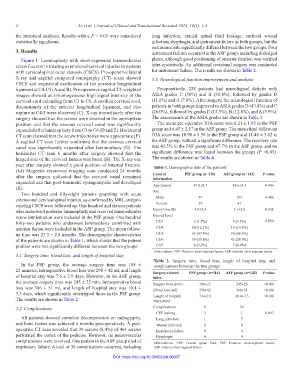
Figure 1. Laminoplasty with short-segmental transpedicular plates, although good positioning of anterior fixation was verified
screw fixation in treating extensional cervical injuries in patients intra-operatively. An additional revisional surgery was conducted
with cervical spinal canal stenosis (CSCS). Pre-operative lateral for instrument failure. The results are shown in Table 2.
X-ray and sagittal computed tomography (CT) scans showed 3.3. Neurological function improvement and analysis
CSCS and segmental ossification of the posterior longitudinal
ligament at C4-C5 (A and B). Pre-operative sagittal T2-weighted Preoperatively, 258 patients had neurological deficits with
images showed an inhomogeneous high signal intensity of the ASIA grades C (50%) and B (31.4%), followed by grades D
cervical cord extending from C3 to C6. A swollen cervical cord, (11.6%) and A (7.0%). After surgery, the neurological function of
discontinuity of the anterior longitudinal ligament, and disc patients in both groups improved to ASIA grades D (43.8%) and C
rupture at C4/5 were observed (C). X-ray immediately after the (26.0%), followed by grades E (13.5%), B (12.8%), and A (3.9%).
surgery showed that the screws were inserted in the appropriate The assessments of the ASIA grades are shown in Table 3.
position and that the osseous cervical canal was significantly The mean pre-operative JOA score was 6.21 ± 1.85 in the PSF
expanded after laminoplasty from C3 to C6 (D and E). Horizontal group and 6.45 ± 2.17 in the ASF group. The mean final follow-up
CT scan showed that the screw trajectories were appropriate (F). JOA score was 10.90 ± 3.56 in the PSF group and 11.48 ± 3.62 in
A sagittal CT scan further confirmed that the osseous cervical the ASF group, without a significant difference. The recovery rate
canal was significantly expanded after laminoplasty (G). The was 43.5% in the PSF group and 47.7% in the ASF group, and no
horizontal CT scan 6 months after surgery showed that the significant difference was found between the groups (P >0.05).
hinged side of the cervical lamina was fused (H). The X-ray one The results are shown in Table 4.
year after surgery showed a good position of internal fixation. Table 1. Demographic data of the patients
(I-J) Magnetic resonance imaging scan conducted 24 months
after the surgery indicated that the cervical canal remained General PSF group (n=116) ASF group (n=142) P‑value
information
expanded and that post-traumatic syringomyelia had developed Age (years) 47.8±8.7 48.6±8.2 0.446
(K).
Two hundred and fifty-eight patients grappling with acute Sex
extensional cervical spinal injuries, as confirmed by MRI, and pre- Male 87 101 0.486
existing CSCS were followed up. One hundred and sixteen patients Female 29 41
who underwent posterior laminoplasty and received transpedicular Injury time (h) 4.9±2.4 5.1±2.9 0.548
screw implantation were included in the PSF group. One hundred Injured level
forty-two patients who underwent laminoplasty combined with C2/3 2 (1.7%) 5 (3.5%) 0.850
anterior fusion were included in the ASF group. The mean follow- C3/4 14 (12.1%) 19 (13.4%)
up time was 23.5 ± 2.8 months. The demographic characteristics C4/5 55 (47.4%) 70 (49.3%)
of the patients are shown in Table 1, which shows that the patient C5/6 39 (33.6%) 41 (28.9%)
profiles were not significantly different between the two groups. C6/7 6 (5.2%) 7 (4.9%)
Abbreviations: PSF: Posterior short-segment fusion, ASF: Anterior short segment fusion
3.1. Surgery time, blood loss, and length of hospital stay
Table 2. Surgery time, blood loss, length of hospital stay, and
In the PSF group, the average surgery time was 188 ± complications between the two groups
23 minutes, intraoperative blood loss was 298 ± 42 ml, and length Surgery‑related PSF group (n=116) ASF group (n=142) P‑value
of hospital stay was 7.6 ± 2.9 days. However, in the ASF group, index
the average surgery time was 245 ± 25 min, intraoperative blood Surgery time (min) 188±23 245±25 <0.001
loss was 366 ± 51 ml, and length of hospital stay was 10.4 ± Blood loss (ml) 298±42 366±51 <0.001
3.3 days, which significantly outstripped those in the PSF group. Length of hospital 7.6±2.9 10.4±3.3 <0.001
The results are shown in Table 2. stay (days)
3.2. Complications Complications 6 24
CSF leaking 3 1 0.043
All patients showed complete decompression on radiography, Lung infection 1 7
and bone fusion was achieved 6 months postoperatively. A post- Wound infection 2 8
operative CT scan revealed that 39 screws (8.4%) of 464 screws Instrument failure 0 4
perforated the cortex of the pedicles. However, no neurovascular Dysphagia 0 4
complications were involved. One patient in the ASF group died of Abbreviations: CSF: Cranial spinal fluid, PSF: Posterior short-segment fusion,
respiratory failure. A total of 30 complications occurred, including ASF: Anterior short segment fusion
DOI: https://doi.org/10.36922/jctr.00037