
Page 68 - MI-1-1
P. 68
Microbes & Immunity Food chain milieus as a reserver for multidrug resistance
2.2. Isolation of bacteria and identification 2.5. Gene detection
Cetrimide selective agar (CSA) supplemented with 10% Polymerase chain reaction (PCR) analysis was used to
glycerol was used for culturing all samples. Incubation detect MBL genes (bla NDM , bla IMP-1 , bla IMP-2 , and bla VIM )
was performed overnight at 37°C. Putative colonies of in the P. aeruginosa isolates that showed reduced
4,12
P. aeruginosa that produced bluish or greenish colonies susceptibility to any of the tested carbapenems (IPM or
(as a result of pigmentation) were recovered and plated on ETP) in accordance with a previously used protocol. 11,14,15
fresh CSA plates to recover pure isolates of Pseudomonas, DNA was extracted from the P. aeruginosa isolates using
which were further subjected to biochemical tests including the GENESpin DNA isolation kit (Eurofins, Hamburg)
oxidase, indole, urease, catalase, and citrate tests for the according to the manufacturer’s guidelines. The extracted
identification of P. aeruginosa strains. 12 DNA was stored at -20°C for the PCR analysis. The
PCR mix was a final volume of 26.5 μl reaction mixture
2.3. Antibiotic susceptibility studies comprising: 0.2 μl of Taq pol U/μl, 2.5 μl of 10X PCR
This experiment was undertaken using our previous protocol buffer, 2.5 μl MgCl , 1μl of 10 pM from each of the forward
2
on the determination of the antibiotic susceptibility of and reverse primers (Table 1), 2.5 μl of dNTPs MIX
bacteria in a non-hospital environment . All P. aeruginosa (2 mM), 3 μl of DNA template, and 14.8 μl of nuclease-
8
isolates were subjected to antibiotic susceptibility studies free water. The presence of MBL genes in the P. aeruginosa
using single antibiotic disks (Oxoid, UK) comprising: isolates was investigated by PCR amplification in a thermal
Fosfomycin (FOS; 10 μg), tetracycline (TE; 10 μg), cefoxitin cycler (Lumex Instruments, Canada) using defined PCR
(FOX; 30 μg), IPM; 10 μg, ceftazidime (30 μg), ertapenem conditions (Table 1). DNA amplicons were run in a 1.5%
(ETP; 30 μg), gentamicin (CN; 10 μg), amikacin (AK; 10 μg), agarose gel electrophoresis for 60 m at 120 V while an
ciprofloxacin (CIP; 10 μg), cefotaxime (CTX; 30 μg), ultraviolet transilluminator (Scientico, India) was used
sulfamethoxazole-trimethoprim (SXT; 25 μg), aztreonam for the visualization of the amplified DNA products in the
(30 μg), and nitrofurantoin (F; 10 μg). The susceptibility tested isolates.
test disks were placed 24 mm apart on freshly prepared 2.6. Plasmid curing studies
Mueller–Hinton (MH) agar plates (Oxoid, UK) inoculated
with the test bacteria (adjusted to 0.5 McFarland’s turbidity Plasmid curing was carried out using acridine orange as
standards). All susceptibility test plates were incubated at the plasmid curing agent. Two colonies of individual MBL-
37°C for 18 – 24 h. The zone diameter of each antibiotic producing P. aeruginosa isolates were grown in 5 ml of
disk was measured and reported as susceptible or resistant nutrient broth supplemented with 0.1 mg/ml of acridine
according to the standard antibiotic breakpoints and orange. Positive control (P. aeruginosa alone) and negative
guidelines of the Clinical Laboratory Standard Institute, control (acridine orange alone) tubes were run alongside
2019. 13 the curing experiments. All tubes were incubated
overnight at 37°C in the incubator. Tubes containing
2.4. MBL
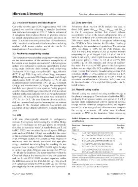
MBL was phenotypically detected in carbapenem- Table 1. Polymerase chain reaction conditions for MBL gene
resistant P. aeruginosa isolates using the modified Hodge amplification
test (MHT) technique on MH agar plates, as previously Resistance Gene sequence Amplicon
described in our earlier study. A standard strain of E. coli gene size (bp)
4
(ATCC 25922) was inoculated by spreading on a MH agar target (s)
plate, and an IPM disk (10 μg) was applied at the center bla NDM Fm (5’-GGTTTGGCGATCTGGTTTTC-3’) 264
of the inoculated MH agar plate. Thereafter, a suspension Rm (5’-CGGAATGGCTCATCACGATC-3’)
of an overnight culture of P. aeruginosa was streaked or bla IMP‑1 F1 (5’-ACC GCA GCA GAG TCT TTG CC-3’) 587
inoculated onto the already inoculated MH agar plate R1 (5’-ACA ACC AGT TTT GCC TTA CC-3’)
using a sterilized inoculating loop by streaking the test bla IMP‑2 F2 (5’-GTT TTA TGT GTA TGC TTC C-3’) 678
organism from the edge of the IPM disk (10 μg) to the edge R2 (5’-AGC CTG TTC CCA TGT AC-3’)
of the MH agar plate. All plates were incubated at 37°C for bla VIM F3 (5’-AGT GGT GAG TAT CCG ACA G-3’) 261
18 – 24 h in the incubator. The plates were observed for the R3 (5’-ATG AAA GTG CGT GGA GAC-3’)
Cloverleaf effect at the intersection of the test bacterium Notes: Amplification conditions: Initial denaturation at 94°C for 3 min,
and the E. coli ATCC 25922 standard organism, within followed by 25 cycles of DNA denaturation at 94°C (30 s); annealing at
the inhibition zone of the IPM disk (10 μg). The growth 64°C (30 s); extension at 72°C (1 min); and final extension at 72°C for
5 min.
of P. aeruginosa toward the IPM disk (10 μg) was inferred Abbreviations: bp: Base pair; F: Forward primer;
phenotypically for MBL production by the MHT protocol. 4 MBL: Metallo-beta-lactamase; R: Reverse primer.
Volume 1 Issue 1 (2024) 62 doi: 10.36922/mi.2319