
Page 136 - MI-2-2
P. 136
Microbes & Immunity Establishment of a novel anti-human CCR8 monoclonal antibody
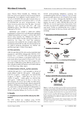
Alum (Thermo Fisher Scientific Inc., Waltham, MA, hCCR8 mAbs-producing hybridoma screening was
USA) was used as an adjuvant for the first immunization. conducted using flow cytometry (Figure 1). Two mice were
Subsequently, three additional weekly injections of 1 × intraperitoneally immunized with LN229/hCCR8 weekly
10 cells of LN229/hCCR8 were administered without an for a total of 5 times. Subsequently, hybridomas were
8
8
adjuvant. A final booster immunization with 1 × 10 cells seeded into 96-well plates, after which flow cytometric
of LN229/hCCR8 was given intraperitoneally 2 days analysis was used to select CHO/hCCR8-reactive and
before harvesting splenocytes from the mice. Harvested CHO-K1-non-reactive supernatants of hybridomas. From
splenocytes were then fused with P3U1 cells using 956 wells, only one (0.10%) yielded CHO/hCCR8-reactive
polyethylene glycol 1500 (PEG1500; Roche Diagnostics, supernatant. Finally, we established the clone C Mab-21
8
Indianapolis, IN, USA). (mouse IgM, kappa) by limiting dilution and additional
screening.
Hybridomas were cultured in RPMI-1640 medium
supplemented as shown above and additional supplements
included hypoxanthine, aminopterin, and thymidine (HAT; A
Thermo Fisher Scientific, Inc., Waltham, MA, USA), 5%
Briclone (NICB, Dublin, Ireland), and 5 μg/mL of Plasmocin
(InvivoGen, San Diego, CA, USA). The hybridoma
supernatants were screened by flow cytometry using CHO/
hCCR8 and parental CHO-K1 cells. The cultured supernatant
of C Mab-21-producing hybridomas was filtrated and
8
purified using Capto L (Cytiva, Tokyo, Japan). B
2.4. Flow cytometry
CHO-K1 and CHO/hCCR8 cells were harvested after brief
exposure to 1 mM EDTA. Subsequently, CHO-K1, CHO/
hCCR8, TALL-1, and CCRF-HSB2 cells were washed with
0.1% bovine serum albumin in phosphate-buffered saline
and treated with primary mAbs for 30 min at 4°C. The cells
were then treated with Alexa Fluor 488-conjugated anti- C
mouse IgG (1:1000) following the collection of fluorescence
data, using the SA3800 Cell Analyzer and SA3800 software
ver. 2.05 (Sony Corp, Tokyo, Japan).
2.5. Determination of the EC by flow cytometry
50
CHO/hCCR8 and TALL-1 were suspended in 100 μL of
serially diluted C Mab-21 (100 μg/mL – 0.006 μg/mL),
8
S19017D (10 μg/mL – 0.0006 μg/mL for CHO/hCCR8; D
10 μg/mL, 2.5 μg/mL – 0.0006 μg/mL for TALL-1), or
L263G8 (10 μg/mL – 0.0006 μg/mL for CHO/hCCR8;
0.625 μg/mL – 0.0006 μg/mL for TALL-1). Alexa Fluor
488-conjugated anti-mouse IgG (1:200) was then added.
Fluorescence data were subsequently collected using the BD
FACSLyric (BD Biosciences, Franklin Lakes, NJ, USA), and
EC values were calculated by fitting the binding isotherms Figure 1. A schematic procedure of anti-hCCR8 monoclonal antibodies
50
into the built-in one-site binding model in GraphPad production. The procedure of the CBIS method for antibody development.
PRISM 6 (GraphPad Software, Inc., La Jolla, CA, USA). LN229/hCCR8 cells were immunized into two mice via intraperitoneal
injection (A). Spleen cells harvested from mice were fused with P3U1
3. Results myeloma cells (B). The culture supernatants of hybridoma were screened
by flow cytometry using CHO-K1 and CHO/hCCR8 (C). After limiting
3.1. Establishment of anti-hCCR8 mAbs by the CBIS dilution of hybridomas and additional analysis, the C Mab-21 clone was
method finally established (D). 8
Abbreviations: i.p.: Intraperitoneal; CHO: Chinese hamster ovary; CBIS:
The CBIS method was employed using hCCR8- Cell-based immunization and screening; hCCR8: Human C-C motif
overexpressing cells to develop anti-hCCR8 mAbs. Anti- chemokine receptor-8.
Volume 2 Issue 2 (2025) 128 doi: 10.36922/mi.4661