
Page 66 - MSAM-3-4
P. 66
Materials Science in Additive Manufacturing 3D printing of anti-microbial parts
A B C
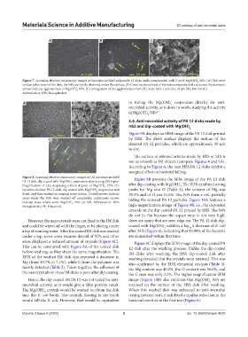
Figure 7. Scanning electron microscopy images of injection-molded polyamide 12 disks melt-compounded with 5 wt.% Mg(OH) NPs. (A) The outer
2
surface (plan view) of the disk; the NPs are faintly observed under the surface. (B) Cross-sectional view of the nanocomposite disk cut across the diameter;
arrows indicate agglomerates of Mg(OH) NPs. (C) enlargement of the agglomerates from (B). Scale bars: 1 µm (A); 10 µm (B); 100 nm (C).
2
Abbreviation: NPs: Nanoplatelets
A B to testing the Mg(OH) suspension directly for anti-
2
microbial activity, as is done in works studying the activity
of Mg(OH) NPs .
37
2
3.4. Anti-microbial activity of PA 12 disks made by
HSS and dip-coated with Mg(OH) 2
Figure 9A displays an SEM image of the PA 12 disk printed
C by HSS. The disk’s surface displays the outline of the
sintered PA 12 particles, which are approximately 50 µm
in size.
The surface of sintered articles made by HSS or SLS is
not as smooth as IM objects (compare Figures 4 and 5A).
According to Figure 6, the neat HSS PA 12 disks exhibit a
marginal effect on bacterial killing.
Figure 8. Scanning electron microscopy images of (A) injection-molded Figure 9B presents the SEM image of the PA 12 disk
PA 12 disk, dip-coated with Mg(OH) suspension after drying; (B) higher
2
magnification of (A); displaying a thick deposit of Mg(OH) NPs; (C) after dip-coating with Mg(OH) . The EDX exhibited strong
2
2
injection-molded PA 12 disk, dip coated with Mg(OH) suspension and peaks for Mg and O (Table 3); the content of Mg was
2
dried, and then washed in running water stream. Dotted arrows indicate 39.3% and of O was 51.6%. The NPs form a veil, partially
areas where the NPs were washed off completely; continuous arrows hiding the sintered PA 12 particles. Figure 10A features a
indicate areas where some Mg(OH) NPs are left. Abbreviation: NPs:
2
Nanoplatelets; PA: Polyamide high-magnification image of Figure 9B, i.e., the deposited
crystals on the dip-coated PA 12 printed by HSS. The NPs
do not lie flat because the aspect ratio is not very high;
However, the nanocrystals were not fixed to the IM disk there are many that are seen edge-on. The PA 12 disk dip-
and could be wiped off with the finger, or by placing under coated with Mg(OH) exhibits a log 4 decrease of E. coli
2
10
a tap of running water. After the coated IM disk was washed after 24 h (Figure 6), indicating that 99.99% of the bacteria
under a tap, some areas became devoid of NPs and other are eliminated within that time.
areas displayed a reduced amount of crystals (Figure 8C). Figure 9C displays the SEM image of the dip-coated PA
This can be contrasted with Figure 8A of the coated disk 12 disk after the washing process. Unlike the dip-coated
before washing, as they share the same magnification. The IM disks after washing, the HSS dip-coated disk after
EDX of the washed IM disk also reported a decrease in washing revealed that the crystals were retained. This was
Mg (from 45.7% to 7.1%), while C from the polymer was also confirmed by the EDX elemental analysis (Table 3);
mostly detected (Table 2). Taken together, the adhesion of the Mg content was 40.2%, the O content was 56.6%, and
the nanocrystals on these IM disks is poor after dip coating. the C seen was only 3.2%. The higher magnification SEM
Hence, the dip-coated IM PA 12 was not tested for anti- image (Figure 10B) also confirms that Mg(OH) NPs are
2
microbial activity, as it would give a false positive result. retained on the surface of the HSS disk after washing.
The Mg(OH) crystals would be washed out from the disk When this washed disk was subjected to anti-microbial
2
into the E. coli broth. The crystals floating in the broth testing (second test), it exhibited a similar reduction in the
would kill the E. coli. However, that would be equivalent bacterial count as in the first test (Figure 6).
Volume 3 Issue 4 (2024) 9 doi: 10.36922/msam.4970