
Page 124 - OR-1-3
P. 124
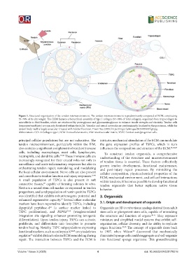
Figure 1. Structural organization of the tendon microenvironment. The tendon microenvironment is predominantly composed of ECM, constituting
            55–70% of its dry weight. This ECM features a hierarchical assembly of type I collagen (97–98% of total collagen), organized from tropocollagen to
            microfibrils to fibril bundles, which are reinforced by proteoglycans and glycosaminoglycans to enhance tensile strength and elasticity. Tendon cells
            (tenocytes/tenoblasts) are sparsely distributed within the ECM. Vascular and neural networks are predominantly localized to the paratenon, while the
            tendon body itself is largely avascular. Created with Adobe Illustrator, Yiwen Xue (2025) https://imgur.la/images/2025/09/09/fig1.jpg.
            Abbreviations: COL I: Collagen type I; ECM: Extracellular matrix; IFM: Interfascicular matrix; TSPC: Tendon stem/progenitor cells.
            principal cellular populations but are not exhaustive. The   intricate; mechanical stimulation of the ECM can modulate
            tendon  microenvironment,  particularly  within  the  IFM,   the gene expression profiles of TSPCs, which in turn
            also contains a significant complement of resident immune   influences the composition and structure of the ECM. 21,24,47
            cells, including macrophages, mast cells, lymphocytes,   To construct tendon organoids, a comprehensive
            neutrophils, and dendritic cells. 37,38  These immune cells are   understanding of the structure and microenvironment
            increasingly recognized for their crucial roles not only in   of tendon tissue is essential. These factors collectively
            surveillance and acute inflammatory responses but also in   govern tendon development, functional maintenance,
            orchestrating tendon repair, remodeling, and modulating   and post-injury repair processes. By mimicking the
            the local cellular environment. Nerve cells are also present   cellular composition, physicochemical properties of the
            and contribute to tendon function and injury responses. 39,40    ECM, mechanical environment, and cell-cell interactions
            A small population of TSPCs is also present in soft   within tendons, it becomes possible to develop functional
            connective tissues,  capable of forming colonies in vitro.   tendon  organoids  that better  replicate  native  tissue
                           29
            Nestin is a neural stem cell marker co-expressed in tendon   behavior.
            progenitors, and a subpopulation of nestin-positive TSPCs
            was identified that exhibits robust tenogenic potential and   3. Organoids
            enhanced regenerative capacity.  Several other molecular
                                      33
            markers have been reported to identify TSPCs, including   3.1. Origin and development of organoids
            dipeptidyl peptidase-4  (a surface marker linked to   Organoids are 3D in vitro tissue analogs derived from adult
                               41
            TSPC  proliferation),  and  RSPO2 42,43   (wingless-related   stem cells or pluripotent stem cells, capable of mimicking
            integration  site  signaling  enhancer  promoting  tenogenic   the structure and function of organs. 48-51  They represent
            differentiation). Upon tendon injury, TSPCs can activate,   miniature and simplified model systems that exhibit self-
            proliferate, and differentiate into tenocytes to promote   organization, cellular diversity, and the ability to replicate
            tendon healing. Notably, TSPC subpopulations expressing   organ functions. 49,50  The concept of organoids dates back
            functional markers, such as cathepsin K 44,45  or cytoskeleton   to 1907, when Wilson  discovered that mechanically
                                                                                  52
            regulator  exhibit distinct roles in ECM remodeling during   dissociated sponge cells could reaggregate and self-organize
                    46
            repair. The interaction between TSPCs and the ECM is   into functional sponge organisms. This groundbreaking


            Volume 1 Issue 3 (2025)                         4                            doi: 10.36922/OR025170016