
Page 55 - TD-1-2
P. 55
Tumor Discovery Adult cystic hyroma
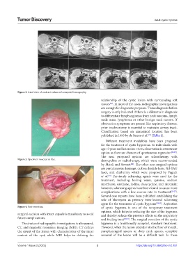
Figure 2. Axial view of contrast-enhanced computed tomography.
relationship of the cystic lesion with surrounding soft
tissues . In most of the cases, radiographic investigations
[21]
are enough for diagnostic purposes. Tissue diagnosis before
surgery is only indicated if there is a dilemma in diagnosis
to differentiate lymphangiomas from neck sarcoma, lymph
node mass, lymphoma or other benign neck tumors. If
obstructive symptoms are present like respiratory distress,
prior tracheostomy is essential to maintain airway track.
Classification based on anatomical location has been
[22]
published in 1995 by de Serres et al. (Table 1).
Different treatment modalities have been proposed
for the treatment of cystic hygromas. In individuals with
age <3 years and lesion size <4 cm, observation is a treatment
option as there are chances of spontaneous regression [22,23] .
The next proposed options are sclerotherapy with
Figure 3. Specimen resected en bloc. doxycycline or radiotherapy, which were recommended
[24]
by Miceli and Stewart . The other non-surgical options
are percutaneous drainage, carbon dioxide laser, Nd-YAG
laser, and diathermy which were proposed by Fageeh
et al. Previously sclerosing agents were used for the
[23]
treatment, including boiling water, quinine, sodium
morrhuate, urethane, iodine, doxycycline, and nitromin;
however, sclerosing agents have been found to cause more
complications with a low success rate in treatment [25-27] .
Several case reports have been published establishing the
role of bleomycin as primary intra-lesional sclerosing
agent for the treatment of cystic hygroma [28,29] . Aspiration
Figure 4. Post-resection. of cystic hygroma is one of the temporary treatment
options, which helps in reducing the size of the hygroma
surgical excision with intact capsule is mandatory to avoid and thereby reduces the pressure effects on the respiratory
future complications. and feeding tract [30,31] . The surgical resection of the cystic
The choice of radiographic investigations is ultrasound, hygroma is a traditionally accepted, standard treatment.
CT, and magnetic resonance imaging (MRI). CT defines However, when the lesion extends into the floor of mouth,
the extent of the lesion with characteristics of the inner parapharyngeal spaces or deep neck spaces, complete
content of the cyst, while MRI helps in defining the removal of the lesion will be a difficult task. In these
Volume 1 Issue 2 (2022) 3 https://doi.org/10.36922/td.v1i2.151