
Page 28 - TD-2-1
P. 28
Tumor Discovery An approach for classification of lung nodules
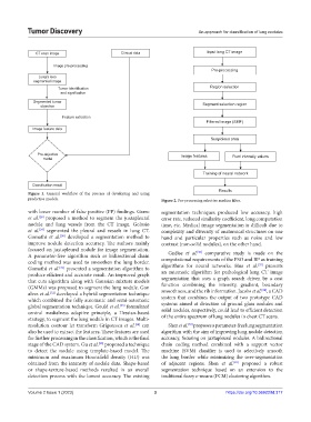
CT scan image Clinical data Input lung CT image
Image pre-processing
Pre-processing
Lung’s less
segmented image
Tumor identification Region selection
and signification
Segmented tumor Segment selection region
objective
Feature extraction
Filtered image (SMF)
Image feature data
Suspicious area
Pre-objective Image features Pixel intensity values
model
Training of neural network
Classification result
Results
Figure 1. General workflow of the process of developing and using
predictive models. Figure 2. Pre-processing selective median filter.
with lesser number of false positive (FP) findings. Garro segmentation techniques produced low accuracy, high
et al. proposed a method to segment the juxtapleural error rate, reduced similarity coefficient, long computation
[28]
nodule and lung vessels from the CT image. Golosio time, etc. Medical image segmentation is difficult due to
et al. segmented the pleural and vessels in lung CT. complexity and diversity of anatomical structures on one
[29]
[30]
Gomathi et al. developed a segmentation method to hand and particular properties such as noise and low
improve nodule detection accuracy. The authors mainly contrast (non-solid nodules), on the other hand.
focused on juxtapleural nodule for image segmentation. [36]
A parameter-free algorithm such as bidirectional chain Gudise et al. comparative study is made on the
coding method was used to smoothen the lung border. computational requirements of the PSO and BP as training
[37]
Gomathi et al. presented a segmentation algorithm to algorithms for neural networks. Hua et al. presents
[31]
produce efficient and accurate result. An improved graph an automatic algorithm for pathological lung CT image
that cuts algorithm along with Gaussian mixture models segmentation that uses a graph search driven by a cost
(GMMs) was proposed to segment the lung nodule. Gon function combining the intensity, gradient, boundary
[38]
[32]
alves et al. developed a hybrid segmentation technique smoothness, and the rib information. Jacobs et al. , a CAD
which combined the fully automatic and semi-automatic system that combines the output of two prototype CAD
systems aimed at detection of ground glass nodules and
global segmentation technique. Gould et al. formulated
[33]
central medialness adaptive principle, a Hessian-based solid nodules, respectively, could lead to efficient detection
strategy, to segment the lung nodule in CT images. Multi- of the entire spectrum of lung nodules in chest CT scans.
resolution contour let transform Grigorescu et al. can Shen et al. proposes a parameter-free lung segmentation
[34]
[39]
also be used to extract the features. These features are used algorithm with the aim of improving lung nodule detection
for further processing in the classification, which is the final accuracy, focusing on juxtapleural nodules. A bidirectional
stage of the CAD system. Gu et al. proposed a technique chain coding method combined with a support vector
[35]
to detect the nodule using template-based model. The machine (SVM) classifier is used to selectively smooth
minimum and maximum Hounsfield density (HU) was the lung border while minimizing the over-segmentation
[40]
obtained from the intensity of nodule data. Shape-based of adjacent regions. Shen et al. proposed a robust
or shape-texture-based methods resulted in an overall segmentation technique based on an extension to the
detection process with the lowest accuracy. The existing traditional fuzzy c-means (FCM) clustering algorithm.
Volume 2 Issue 1 (2023) 3 https://doi.org/10.36922/td.317