
Page 119 - TD-3-1
P. 119
Tumor Discovery Inflammatory orbital pseudotumors
CD20 monoclonal antibodies (rituximab 375 mg/m ). level of 85 mg/L. Renal function tests yielded normal
2
A subsequent PET scan after 1 month of rituximab therapy results, as did the thyroid test. Furthermore, serological
demonstrated a decrease in metabolic activity (Figure 2D). tests indicated a history of successfully treated chronic
A radiological follow-up was planned after 6 months of hepatitis B with an undetectable viral load. Serological tests
treatment, with maintenance therapy using rituximab. for hepatitis C, human immunodeficiency virus, syphilis,
and toxoplasmosis returned negative results. In addition,
2.3. Case 3 the tuberculosis screening was negative.
A 58-year-old man with a history of bilateral cataracts for The bone marrow biopsy revealed high cellularity
1 year presented with an alteration in his general condition. indicative of a regenerating marrow. However,
General examination revealed an anemic syndrome, immunohistochemistry results (CD20, CD3, CD34,
splenomegaly with a 6 cm costal margin, hepatomegaly, CD15, CD68, and CD30) were inconclusive, failing
a solid, and non-pulsatile left orbital tumor (Figure 3A), to definitely rule out a tumoral origin. Subsequently,
accompanied by conjunctival jaundice. Orbital MRI revealed the patient was initiated on corticosteroid therapy at a
a well-defined oval lesion formation in the left palpebral dose of 1 mg/kg/day. Based on the remarkable clinical
region with regular hyposignal contours on T1 and T2 improvement observed (Figure 3B), including weight
sequences, without extension into the end orbit (Figure 3C). regain, alleviation of asthenia, and resolution of the orbital
In addition, scans indicated hepatosplenomegaly measuring mass, a diagnosis of inflammatory orbital pseudotumor
24 cm, with the spleen also measuring 24 cm. with associated hemolytic anemia was established. Notably,
Biological findings revealed profound anemia, normalization of abnormal blood counts and resolution
characterized by a hemoglobin level of 4 g/dl. In of the inflammatory syndrome were observed as early as
addition, leukopenia was observed with a leukocyte 3 weeks into treatment. Follow-up imaging at 2 months
count of 2264/mm , along with lymphopenia revealed partial remission of the condition (Figure 3D),
3
(lymphocyte count at 765/mm ) and thrombocytopenia while scans at 6 months indicated complete resolution of
3
(platelet count at 9000/mm ). A regenerative reticulocyte hepatomegaly and splenomegaly. Although a palpebral
3
count was noted, along with a positive indirect Coombs biopsy was proposed, the patient was lost to follow-up.
test. The phospho-calcium balance was within normal 3. Discussion
limits. Inflammatory markers showed a positive polyclonal
profile on protein electrophoresis, with a C-reactive protein Inflammatory diseases of the orbit remain poorly defined
in terms of etiology and pathogenesis. Various factors
A B such as infection, immune response, post-traumatic
conditions, or molecular mimicry have been proposed
as potential causes. The administration of steroids and
1
immunosuppressants is often considered favorable, given
the observed increase in inflammation cytokines.
It is important to note the potential diagnostic challenges
C D posed by persistent and progressive tumors resistant to
corticosteroid treatment, as observed in Case 1. Such cases
raise concerns regarding the presence of an underlying
aggressive neoplastic pathology. Bilateralization of the
9
inflammatory process, occurring in 8 – 20% of cases, and
10
exaggerated edema and incoercible periorbital inflammation,
led to exenteration. To the best of our knowledge, Case 1
represents the first case of an orbital pseudotumor revealing
Takayasu vasculitis of the large trunk.
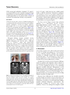
Figure 3. Clinical presentations of Case 3. (A) External photograph of
the patient displaying exophthalmos, which developed over 3 months However, Case 2 illustrates the favorable (essentially
along with hemolytic anemia and tumor syndrome. (B) Complete clinical) evolution of idiopathic pseudo-orbital tumors, as
11
clinical improvement observed. (C) Axial scan revealing a well-defined observed in 70% of cases under corticosteroid treatment.
oval-shaped lesion in the left upper palpebral region, exhibiting hypo Nonetheless, the persistence of radiological abnormalities
T1 and T2 signals with mild contrast enhancement. (D) Treatment with required additional therapeutic measurements. The
corticosteroids resulted in partial regression of the left upper palpebral
mass, as observed in the hyposignal T2, with slight enhancement after management strategies for refractory cases remain
contrast administration in the axial sequence. undefined. However, the etiological investigation should
Volume 3 Issue 1 (2024) 4 https://doi.org/10.36922/td.1792