
Page 52 - TD-3-2
P. 52
Tumor Discovery Schwannoma misdiagnosis risks
region behind the mandibular angle. The patient exhibited
no signs of numbness, compression (such as dysphagia
or dyspnea), or discomfort. On examination, a firm
painless mass, approximately 2 cm × 2 cm, was palpable
at level 2 on the right side. The mass was mobile in both
superficial and deep planes, with no skin changes overlying.
Oto-rhino-laryngoscopic examination revealed normal
findings. Ultrasound examination identified a well-defined
solid cystic mass, measuring 2.5 cm in diameter, hypoechoic
with no internal flow. There was no continuity between the
proximal and the distal ends. Infracentimetric lymph node
structures were identified alongside the mass. Given their
proximity, the original diagnosis of pathological lymph
node was maintained, and thus, no further radiological
exploration was pursued. A fine-needle aspiration cytology
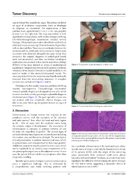
(FNAC) of the mass showed an atypia of undetermined Figure 1. Peroperative image showing the nodular mass.
significance. Surgical exploration under general anesthesia
revealed a white nodular formation located medially to the
anterior border of the sternocleidomastoid muscle. The
mass was distant from the neurovascular bundle and easily
dissected from the surrounding structures. A complete
excision was carried out (Figures 1 and 2).
Histologically, the nodular mass was calcified, exhibiting
necrotic rearrangement. Histopathologic examination
revealed spindle-shaped and elongated tumor cells, which
showed no mitotic activity, pointing to a plausible diagnosis
of schwannoma (Figure 3). The post-operative course was
not marked with any remarkable clinical changes, and
after a one-year follow-up, the patient showed no signs of
recurrence. Figure 2. Excised mass before histological examination.
3. Discussion
A B
Schwannomas are benign tumors that originate in any
peripheral nerves, with the exception of the olfactory
and optic nerves. They affect the head-and-neck region
4
in 20 – 45% of cases, with the vestibular nerve being
the primary origin of development. The occurrence of
5
schwannomas is common in patients between 20 and
50 years old, regardless of gender. The clinical signs of
4
cervical schwannomas depend on various factors including Figure 3. Microscopic examination of hematoxylin-eosin-stained sections
of surgically resected tumor under ×20 (A) and ×40 (B) magnification.
the location, size, and the nerve of origin. The most typical The sections show cytologically bland spindle cells against a vague nuclear
clinical presentation is isolated laterocervical mass, which palisading and fibrillary background.
is asymptomatic and characterized by slow mass growth.
5
Additional symptoms may be present in some cases, which non-vestibular schwannomas of the head and neck, where
can be explained by an external mass compression of the specific nerve of origin could only be determined in 16 out
oro-pharyngolaryngeal axes or the adjacent nerves. In our of 26 patients (62%), with the origin of the remaining
4
case, the patient did not exhibit any symptoms other than cases, presumably from unnamed small nerve plexus
progressive swelling. branches, left unidentified. Similarly, an article from India
4
Identifying the nerve of origin in cervical schwannomas highlights the difficulty of determining the nerve of origin
poses a significant challenge. This is evidenced by for this tumor entity, suggesting a successful chance of
a comprehensive case study involving patients with identification as low as 50%. 3
Volume 3 Issue 2 (2024) 2 doi: 10.36922/td.2606