
Page 56 - TD-3-2
P. 56
Tumor Discovery High-grade sinonasal adenocarcinoma
mortality rates in contrast to alternative surgical
procedures. Radiotherapy pre-dominantly serves as a
5,6
palliative or complementary measure in the post-operative
phase. Despite its positive effects, chemotherapy has
5,7
waned in popularity due to its suboptimal reproducibility
of therapeutic effects. Evidently, surgery remains the
most advantageous treatment modality, whether utilized
independently or in conjunction with radiotherapy.
5
Adjuvant radiotherapy is recommended for high-grade
tumors and those classified as T3 or T4 stage. 6
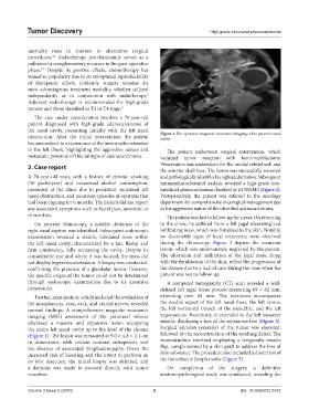
The case under consideration involves a 70-year-old
patient diagnosed with high-grade adenocarcinoma of
the nasal cavity, presenting initially with the left nasal
obstruction. After the initial intervention, the patient Figure 1. Pre-operative magnetic resonance imaging of the patient’s nasal
cavity
became subject to a recurrence of the tumor with extension
to the left cheek, highlighting the aggressive nature and The patient underwent surgical intervention, which
metastatic potential of this subtype of adenocarcinoma. included tumor resection with hemi-maxillectomy.
2. Case report Preservation was undertaken for the medial orbital wall and
the anterior skull base. The lesion was successfully removed
A 70-year-old man, with a history of chronic smoking and pathologically identified as a glandular tumor. Subsequent
(30 packs/year) and occasional alcohol consumption, immunohistochemical analysis revealed a high-grade non-
presented at the clinic due to persistent, unilateral left intestinal adenocarcinoma classified as pT3N0M0 (Figure 2).
nasal obstruction, and recurrent episodes of epistaxis that Postoperatively, the patient was referred to the oncology
had been ongoing for 6 months. The patient did not report department for comprehensive oncological management due
any associated symptoms such as facial pain, anosmia, or to the aggressive nature of the identified adenocarcinoma.
rhinorrhea. The patient was lost to follow-up for a year. On returning
On anterior rhinoscopy, a notable deviation of the to the clinic, he suffered from a left jugal ulcerating and
right nasal septum was identified. Subsequent endoscopic infiltrating mass, which was fistulized to the skin. Notably,
examination revealed a sizable, lobulated mass within no discernible signs of local recurrence were observed
the left nasal cavity, characterized by a tan, fleshy, and during the rhinoscopy. Figure 3 depicts the recurrent
firm consistency, fully occupying the cavity. Despite its lesion, which was unfortunately neglected by the patient.
considerable size and where it was located, the mass did The ulceration and infiltration of the jugal mass, along
not display hypervascularization. A biopsy was conducted, with the fistulization of the skin, reflect the progression of
confirming the presence of a glandular tumor. However, the disease due to a lack of care during the time when the
the specific origin of the tumor could not be determined patient was lost to follow-up.
through endoscopic examination due to its extensive A computed tomography (CT) scan revealed a well-
dimensions. defined left jugal tissue process measuring 69 × 62 mm,
Further, examination, which included the evaluation of extending over 60 mm. The extension encompasses
the nasopharynx, eyes, neck, and cranial nerves, revealed the medial aspect of the left nasal fossa, the left ramus,
normal findings. A comprehensive magnetic resonance the left horizontal branch of the mandible, and the left
imaging (MRI) assessment of the paranasal sinuses zygomaticus. Posteriorly, it extended to the left masseter
disclosed a massive and expansive lesion occupying muscle, displaying a loss of the separation line (Figure 4).
the entire left nasal cavity up to the level of the choana Surgical excision (exeresis) of the tumor was executed,
(Figure 1). The lesion was measured at 9.0 × 4.8 × 2.1 cm followed by the reconstruction of the resulting defect. The
in dimensions, with evident contrast entrapment, and reconstruction involved employing a temporalis muscle
the absence of associated lymphadenopathy. Given the flap, complemented by a skin graft to address the loss of
increased risk of bleeding and the intent to perform an skin substance. The procedure also included a dissection of
en bloc resection, the initial biopsy was deferred, and the homolateral lymph nodes (Figure 5).
a decision was made to proceed directly with tumor On completion of the surgery, a definitive
resection. anatomopathological study was conducted, revealing the
Volume 3 Issue 2 (2024) 2 doi: 10.36922/td.2423