
Page 58 - TD-3-2
P. 58
Tumor Discovery High-grade sinonasal adenocarcinoma
Adenocarcinomas detected in their early stage can be
managed with only surgical treatment, which results in an
impressive 5-year survival rate of 83.4%. For patients with
advanced-stage disease, the combined approach of surgery
and radiotherapy, which yields a 5-year survival rate of
66.6%, is recommended. Notably, radiotherapy alone does
not confer a significant benefit in terms of 5-year survival
when compared with no-treatment. 14
This case provides valuable insights, emphasizing the
rarity and aggressiveness of high-grade non-intestinal
sinus adenocarcinomas. Nasal congestion emerges as a
prominent and non-specific symptom, often contributing
to delayed diagnoses. Thus, multidisciplinary assessments
play a pivotal role in effective patient management.
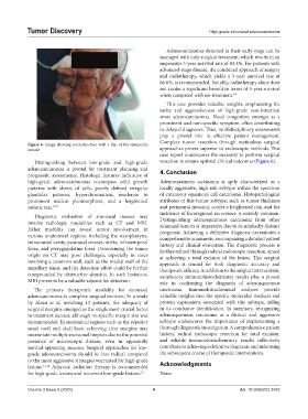
Complete tumor resection through meticulous surgical
Figure 6. Image showing reconstruction with a flap of the temporalis
muscle approaches proves superior to endoscopic methods. This
case report underscores the necessity to perform surgical
Distinguishing between low-grade and high-grade resection to ensure optimal clinical outcomes (Figure 6).
adenocarcinomas is pivotal for treatment planning and 4. Conclusion
prognostic assessments. Histologic features indicative of
high-grade adenocarcinomas encompass solid growth Adenosquamous carcinoma is aptly characterized as a
patterns with sheets of cells, poorly defined irregular locally aggressive, high-risk subtype within the spectrum
glandular patterns, hyperchromatism, moderate to of cutaneous squamous cell carcinoma. Histopathological
prominent nuclear pleomorphism, and a heightened attributes of this tumor subtype, such as tumor thickness
mitotic rate. 9,10 and perineural invasion, confer a heightened risk, and the
incidence of locoregional recurrence is notably common.
Diagnostic evaluation of sinonasal masses may
involve radiologic modalities such as CT and MRI. Distinguishing adenosquamous carcinoma from other
sinonasal tumors is imperative due to its markedly distinct
Either modality can reveal tumor involvement in prognosis. Achieving a definitive diagnosis necessitates a
various anatomical regions, including the nasopharynx, comprehensive anamnesis, encompassing a detailed patient
intracranial cavity, paranasal sinuses, orbits, infratemporal history and clinical evaluation. The diagnostic process is
fossa, and pterygopalatine fossa. Determining the tumor further refined through radical endoscopic resection, aimed
origin on CT may pose challenges, especially in cases at achieving a total excision of the lesion. This surgical
involving a common wall, such as the medial wall of the approach is crucial for both diagnostic accuracy and
maxillary sinus, and the detection effort could be further therapeutic efficacy. In addition to the surgical intervention,
compounded by obstructive sinusitis. In such instances, satisfactory immunohistochemistry results play a pivotal
MRI proves to be a valuable adjunct for detection. role in confirming the diagnosis of adenosquamous
The primary therapeutic modality for sinonasal carcinoma. Immunohistochemical analyses provide
adenocarcinoma is complete surgical excision. In a study valuable insights into the specific molecular markers and
by Alessi et al. involving 13 patients, the adequacy of protein expressions associated with this subtype, aiding
surgical margins emerged as the single most crucial factor in its conclusive identification. In summary, recognizing
in treatment success, although no specific margin size was adenosquamous carcinoma as a distinct and aggressive
recommended. In anatomical regions such as the superior subtype underscores the importance of implementing a
nasal vault and skull base, achieving clear margins may thorough diagnostic investigation. A comprehensive patient
necessitate multiple excisional biopsies due to the potential history, radical endoscopic resection for total excision,
presence of microscopic disease, even in apparently and reliable immunohistochemistry results collectively
normal-appearing mucosa. Surgical approaches for low- contribute to achieving a definitive diagnosis and informing
grade adenocarcinoma should be less radical compared the subsequent course of therapeutic interventions.
to the more aggressive strategies warranted for high-grade
lesions. 11,12 Adjuvant radiation therapy is recommended Acknowledgments
for high-grade lesions and recurrent low-grade lesions. 13 None.
Volume 3 Issue 2 (2024) 4 doi: 10.36922/td.2423