
Page 121 - TD-3-3
P. 121
Tumor Discovery CRMO presenting as multifocal bone LCH
A B
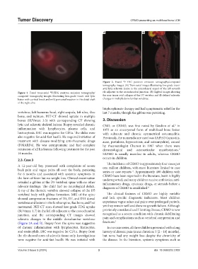
Figure 2. Fused F-FDG positron emission tomography/computed
18
tomography images. (A) Trans-axial images illustrating low-grade tracer
avid lytic-sclerotic lesion in the anterolateral aspect of the left seventh
Figure 1. Fused trans-axial F-FDG positron emission tomography/ rib adjacent to the costochondral junction. (B) Sagittal images showing
18
computed tomography images illustrating low-grade tracer avid lytic the non-tracer avid collapse of the D7 vertebra and ill-defined sclerotic
lesion with cortical break and mild periosteal reaction in the distal shaft changes in multiple dorsolumbar vertebrae.
of the right ulna
bisphosphonate therapy and had symptomatic relief for the
vertebrae, left humerus head, right scapula, left ulna, iliac last 7 months, though the gibbus was persisting.
bone, and ischium. PET-CT showed uptake in multiple
bones (SUVmax: 3.5) with corresponding CT showing 3. Discussion
lytic and sclerotic skeletal lesions. Biopsy revealed chronic CNO, or CRMO, was first noted by Giedion et al. in
6
inflammation with lymphocytes, plasma cells, and 1972 as an aneurysmal form of multifocal bone lesion
histiocytosis. IHC was negative for CD1a. The slides were with subacute and chronic symmetrical osteomyelitis.
also negative for acid-fast bacilli. He required initiation of Previously, the nomenclature used was SAPHO (synovitis,
treatment with disease-modifying anti-rheumatic drugs acne, pustulosis, hyperostosis, and osteomyelitis), coined
(DMARDs). He was asymptomatic and had complete by rheumatologist Chamot in 1987 when there were
remission of all his lesions following treatment for the past dermatological and osteoarticular manifestations.
7
16 months. SAPHO is usually manifest in adults, whereas CRMO
occurs in children.
2.3. Case 3
The incidence of CRMO is approximately four cases per
A 12-year-old boy presented with complaints of severe one million children, with most literature limited to case
back pain and vague pains all over the body, persisting series or case reports . Approximately 400 children with
2
for 6 months and associated with systemic symptoms in CRMO have been reported in the literature, but it is highly
the form of fever but no weight loss. Clinical examination underreported, and many children receive antibiotics, anti-
revealed a gibbus at the D5 vertebral spine with no other inflammatory drugs, cytotoxic drugs, or steroids before a
relevant findings. The child had no neurological deficit. diagnosis of CRMO is established. 8
X-ray of the thoracic vertebra showed collapse of the D5
vertebral body with gibbus formation. MRI of the spine The clinical features of CRMO are highly variable
showed compression fractures of D5, D7, and D11 dorsal and lack specific diagnostic indicators. Most children
vertebrae and lesions in the lumbar spine, iliac bone, and first experience vague aches and pains over prolonged periods,
metatarsal. PET-CT scan showed low-grade FDG uptake yet they remain well and show no growth failure. Although
(SUVmax: 1.7) in the left rib adjacent to the costochondral previously considered a self-limiting disease, CRMO is now
junction, and the corresponding CT images showed recognized as a severe condition with chronic debilitating
sclerotic changes in the middle dorsolumbar vertebrae pain and complications such as vertebral compression and
(Figure 2A and B). Biopsy from the spine was suggestive fractures. 2
of chronic inflammation with lymphocytes, histiocytes, In our case series, all three children presented with a long
and eosinophils. IHC was negative for CD1a. Biopsy from history of chronic pain (mean duration: 5 [2 – 10] months),
the rib showed cores of sclerotic tissue only. Investigations but none had any weight loss despite the chronicity of
were negative for acid-fast bacilli. He was initiated with the disease. In the literature, systemic symptoms such as
Volume 3 Issue 3 (2024) 3 doi: 10.36922/td.3102