
Page 123 - TD-4-1
P. 123
Tumor Discovery Pyrotinib and capecitabine in HER2-negative recurrence
A B C
D E F
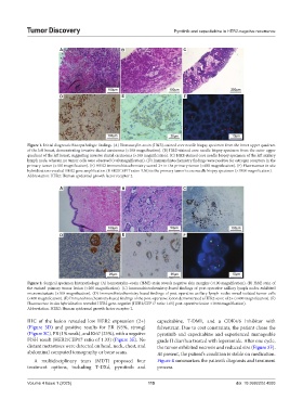
Figure 1. Initial diagnostic histopathologic findings. (A) Hematoxylin-eosin (H&E)-stained core needle biopsy specimen from the inner upper quadrant
of the left breast, demonstrating invasive ductal carcinoma (×100 magnification). (B) H&E-stained core needle biopsy specimen from the outer upper
quadrant of the left breast, suggesting invasive ductal carcinoma (×100 magnification). (C) H&E-stained core needle biopsy specimen of the left axillary
lymph node, wherein no tumor cells were observed (×40 magnification). (D) Immunohistochemistry findings were positive for estrogen receptors in the
primary tumor (×100 magnification). (E) HER2 immunohistochemistry scored 2+ in the primary tumor (×400 magnification). (F) Fluorescence in situ
hybridization revealed HER2 gene amplification (HER2/CEP17 ratio: 3.56) in the primary tumor’s core needle biopsy specimen (×1000 magnification).
Abbreviation: HER2: Human epidermal growth factor receptor 2.
A B C
D E F
Figure 2. Surgical specimen histopathology. (A) hematoxylin–eosin (H&E) stain reveals negative skin margins (×100 magnification). (B) H&E stain of
the excised primary tumor lesion (×200 magnification). (C) Immunohistochemistry-based findings of post-operative axillary lymph nodes exhibited
micrometastasis (×100 magnification). (D) Immunohistochemistry-based findings of post-operative axillary lymph nodes reveal isolated tumor cells
(×400 magnification). (E) Immunohistochemistry-based findings of the post-operative lesion demonstrated a HER2 score of 2+ (×400 magnification). (F)
Fluorescence in situ hybridization revealed HER2 gene-negative (HER2/CEP17 ratio: 1.65) post-operative lesion ×1000 magnification).
Abbreviation: HER2: Human epidermal growth factor receptor 2.
IHC of the lesion revealed low HER2 expression (2+) capecitabine, T-DM1, and a CDK4/6 inhibitor with
(Figure 3D) and positive results for ER (95%, strong) fulvestrant. Due to cost constraints, the patient chose the
(Figure 3C), PR (1% weak), and Ki67 (25%), with a negative pyrotinib and capecitabine and experienced manageable
FISH result (HER2/CEP17 ratio of 1.33) (Figure 3E). No grade II diarrhea treated with loperamide. After one cycle,
distant metastases were detected on head, neck, chest, and the tumor exhibited necrosis and reduced size (Figure 3F).
abdominal computed tomography or bone scans. At present, the patient’s condition is stable on medication.
A multidisciplinary team (MDT) proposed four Figure 4 summarizes the patient’s diagnosis and treatment
treatment options, including T-DXd, pyrotinib and process.
Volume 4 Issue 1 (2025) 115 doi: 10.36922/td.4093