
Page 25 - TD-4-1
P. 25
Tumor Discovery Immunohistochemistry profiling of ovarian cysts
triggered by parthenogenetic processes in younger women. (proteins) of interest in cytology specimens, offering
Gross examination indicates that they are thick-walled, detailed cellular and molecular information on tissues.
8
opaque, and whitish (Figure 4). The contents of dermoid This technique is particularly relevant in diagnostic
cysts include hair, cartilage, bone, and a significant amount cytopathology, offering improved sensitivity and
of sebaceous greasy fluid. Histomorphologically, these specificity for detecting benign and malignant lesions
cysts present with thick walls composed of ectodermal, compared to traditional hematoxylin and eosin staining.
mesodermal, and endodermal tissues. 7,14 ICC can be performed using either direct or indirect
methods, with the latter requiring a secondary antibody
5. Malignant ovarian cysts coupling. According to Kirbiš et al., ICC is considered an
24
Malignant ovarian cysts are less common and primarily invaluable diagnostic technique for establishing empirical
of histological origin, with various subtypes, as shown in diagnosis, predicting biomarkers and prognosis, and
8,24
Figure 5. Epithelial malignant ovarian cysts are the most determining the origin of tumors. It can be applied to
prevalent, accounting for 90% of total cases. The incidence various cytology preparations, including direct smears,
of these cysts is particularly high among postmenopausal liquid-based preparations, cell blocks, cytospins, and cell
women, especially those aged 60 – 70 years. Malignant cultures. Many institutions prefer using cell blocks due
14
ovarian cysts include epithelial ovarian cancer, stromal to their advantage of producing several identical sections
tumors, and germ cell tumors. 13,20 Epithelial ovarian for cases requiring a panel of ICC stains. In addition,
malignant lesions are subdivided into serous carcinoma, most biomarkers have been validated on formalin-fixed
mucinous carcinoma, endometrioid, and clear cell paraffin-embedded (FFPE) tissue, eliminating the need
carcinoma. Malignant germ cell tumors encompass for separate validation. 25-27 With the discovery of specific
embryonal carcinoma, immature teratoma, polyembryoma, biomarkers, ICC has demonstrated the capability to
and endodermal sinus tumors. Risk factors for malignancy distinguish between benign and malignant ovarian cysts. 28
include age, family history, genetic predispositions (such as
BRCA mutations), and certain reproductive histories. 20,21 7. Principles and application of ICC
Histopathologically, malignant ovarian cysts exhibit complex ICC relies on the complementary binding of antibodies to
papillae, necrotic foci, cystic spaces, neuroepithelial cells in target proteins, known as antigens, in cells from pathological
solid sheets, and Call-Exner bodies. 22 specimens. This technique involves labeling antibodies with
6. ICC enzymes, optionally counterstained with hematoxylin and
eosin, and visualizing the results using a light microscope.
ICC is a diagnostic technique that involves applying ICC merges principles from immunology, cytochemistry,
antibodies to detect and visualize cellular antigens and histology to identify specific cellular structures.
The fundamental principle of ICC is the identification,
visualization, and localization of specific antigens based
on a satisfactory signal-to-noise ratio. Signal amplification
and the reduction of non-specific background staining
are crucial strategies for achieving reliable results, which
are invaluable for routine practices. The reaction involves
primary and, in some cases, secondary antibodies, blockers,
enzymes, and enzyme or fluorescent labels. An antibody
29
is an immunoglobulin molecule produced in response
to the presence of an antigen. The choice of antibody for
13
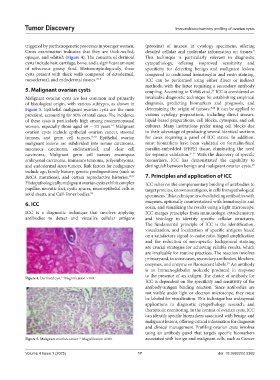
Figure 4. Dermoid cyst. Magnification ×100.
ICC is dependent on the specificity and sensitivity of the
antibody-antigen binding reaction. Since antibodies are
not visible under light or electron microscope, they must
be labeled for visualization. This technique has widespread
applications in diagnostic cytopathology, research, and
therapeutic monitoring. In the context of ovarian cysts, ICC
can identify specific biomarkers associated with benign and
malignant lesions, offering critical information for diagnosis
and clinical management. Profiling ovarian cysts involves
using an antibody panel that targets specific biomarkers
Figure 5. Malignant ovarian cancer. Magnification ×100. associated with benign and malignant cells, such as Cancer
13
Volume 4 Issue 1 (2025) 17 doi: 10.36922/td.5369