
Page 21 - manuscript_ijb05580
P. 21
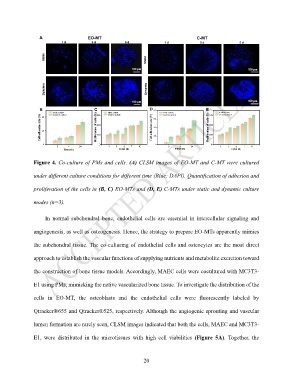
Figure 4. Co-culture of PMs and cells. (A) CLSM images of EO-MT and C-MT were cultured

               under different culture conditions for different time (Blue: DAPI). Quantification of adhesion and

               proliferation of the cells in (B, C) EO-MTs and (D, E) C-MTs under static and dynamic culture


               modes (n=3).


                    In  normal  subchondral  bone,  endothelial  cells  are  essential  in  intercellular  signaling  and


               angiogenesis, as well as osteogenesis. Hence, the strategy to prepare EO-MTs apparently mimics

               the subchondral tissue. The co-culturing of endothelial cells and osteocytes are the most direct

               approach to establish the vascular functions of supplying nutrients and metabolite excretion toward


               the construction of bone tissue models. Accordingly, MAEC cells were cocultured with MC3T3-

               E1 using PMs, mimicking the native vascularized bone tissue. To investigate the distribution of the


               cells  in  EO-MT,  the  osteoblasts  and  the  endothelial  cells  were  fluorescently  labeled  by

               Qtracker®655 and Qtracker®525, respectively. Although the angiogenic sprouting and vascular


               lumen formation are rarely seen, CLSM images indicated that both the cells, MAEC and MC3T3-

               E1,  were  distributed  in  the  microtissues  with  high  cell  viabilities  (Figure  5A).  Together,  the


                                                             20