
Page 25 - manuscript_ijb05580
P. 25
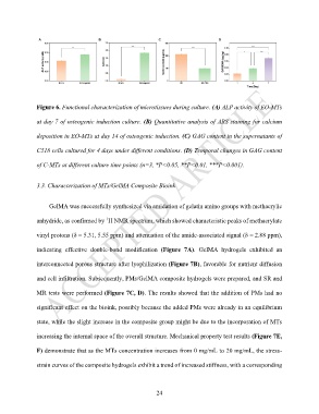
Figure 6. Functional characterization of microtissues during culture. (A) ALP activity of EO-MTs

               at day 7 of osteogenic induction culture. (B) Quantitative analysis of ARS staining for calcium


               deposition in EO-MTs at day 14 of osteogenic induction. (C) GAG content in the supernatants of

               C518 cells cultured for 4 days under different conditions. (D) Temporal changes in GAG content


               of C-MTs at different culture time points (n=3, *P<0.05, **P<0.01, ***P<0.001).


               3.3. Characterization of MTs/GelMA Composite Bioink


                    GelMA was successfully synthesized via amidation of gelatin amino groups with methacrylic


                                          1
               anhydride, as confirmed by  H NMR spectrum, which showed characteristic peaks of methacrylate
               vinyl protons (δ = 5.31, 5.55 ppm) and attenuation of the amide-associated signal (δ = 2.88 ppm),


               indicating  effective  double-bond  modification  (Figure  7A).  GelMA  hydrogels  exhibited  an

               interconnected porous structure after lyophilization (Figure 7B), favorable for nutrient diffusion


               and cell infiltration. Subsequently, PMs/GelMA composite hydrogels were prepared, and SR and

               MR tests were performed (Figure 7C, D). The results showed that the addition of PMs had no


               significant effect on the bioink, possibly because the added PMs were already in an equilibrium

               state, while the slight increase in the composite group might be due to the incorporation of MTs


               increasing the internal space of the overall structure. Mechanical property test results (Figure 7E,

               F) demonstrate that as the MTs concentration increases from 0 mg/mL to 20 mg/mL, the stress-

               strain curves of the composite hydrogels exhibit a trend of increased stiffness, with a corresponding


                                                             24