
Page 27 - manuscript_ijb05580
P. 27
1
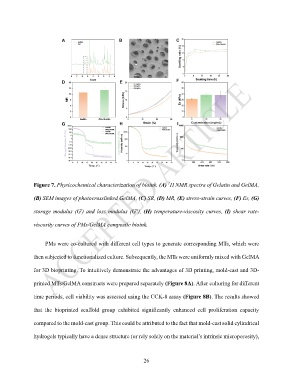
Figure 7. Physicochemical characterization of bioink. (A) H NMR spectra of Gelatin and GelMA,
(B) SEM images of photocrosslinked GelMA, (C) SR, (D) MR, (E) stress-strain curves, (F) Es, (G)
storage modulus (G') and loss modulus (G''), (H) temperature-viscosity curves, (I) shear rate-
viscosity curves of PMs/GelMA composite bioink.
PMs were co-cultured with different cell types to generate corresponding MTs, which were
then subjected to functionalized culture. Subsequently, the MTs were uniformly mixed with GelMA
for 3D bioprinting. To intuitively demonstrate the advantages of 3D printing, mold-cast and 3D-
printed MTs/GelMA constructs were prepared separately (Figure 8A). After culturing for different
time periods, cell viability was assessed using the CCK-8 assay (Figure 8B). The results showed
that the bioprinted scaffold group exhibited significantly enhanced cell proliferation capacity
compared to the mold-cast group. This could be attributed to the fact that mold-cast solid cylindrical
hydrogels typically have a dense structure (or rely solely on the material’s intrinsic microporosity),
26