
Page 23 - manuscript_ijb05580
P. 23
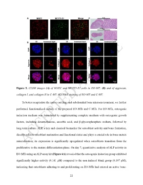
Figure 5. CLSM images (A) of MAEC and MC3T3-E1 cells in EO-MT; (B) and of aggrecan,


               collagen I, and collagen II in C-MT. (C) H&E staining of EO-MT and C-MT.


                    To better recapitulate the native cartilage and subchondral bone microenvironment, we further

               performed functionalized culture of the prepared EO-MTs and C-MTs. For EO-MTs, osteogenic


               induction medium was formulated by supplementing complete medium with osteogenic growth

               factors,  including  dexamethasone,  ascorbic  acid,  and  β-glycerophosphate  sodium,  followed  by


               long-term culture. ALP, a key and classical biomarker for osteoblast activity and bone formation,

               directly reflects osteoblast maturation and functional status and plays a crucial role in bone matrix


               mineralization;  its  expression  is  significantly  upregulated  when  osteoblasts  transition  from  the

               proliferative to the mature differentiation phase. On day 7, quantitative analysis of ALP activity in


               EO-MTs using an ALP assay kit (Figure 6A) revealed that the osteogenic induction group exhibited

               significantly higher activity (0.141 μM) compared to the non-induced blank group (0.107 μM),

               indicating that osteoblasts adhering to and proliferating on EO-MTs had entered an active bone-


                                                             22