
Page 24 - manuscript_ijb05590
P. 24
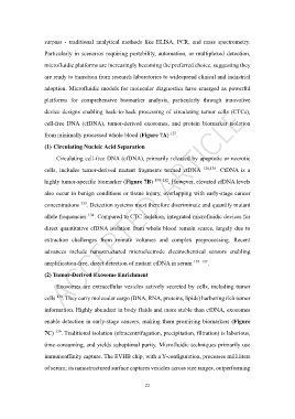
surpass  -  traditional  analytical  methods  like  ELISA,  PCR,  and  mass  spectrometry.

                   Particularly in scenarios requiring portability, automation, or multiplexed detection,

                   microfluidic platforms are increasingly becoming the preferred choice, suggesting they
                   are ready to transition from research laboratories to widespread clinical and industrial

                   adoption. Microfluidic models for molecular diagnostics have emerged as powerful

                   platforms  for  comprehensive  biomarker  analysis,  particularly  through  innovative

                   device designs enabling back-to-back processing of circulating tumor cells (CTCs),

                   cell-free  DNA  (cfDNA),  tumor-derived  exosomes,  and  protein  biomarker  isolation

                   from minimally processed whole blood (Figure 7A)   127 .

                   (1) Circulating Nucleic Acid Separation

                        Circulating cell-free DNA (cfDNA), primarily released by apoptotic or necrotic

                   cells,  includes  tumor-derived  mutant  fragments  termed  ctDNA   128,129 .  CtDNA  is  a

                   highly tumor-specific biomarker (Figure 7B)  130–132 . However, elevated cfDNA levels

                   also occur in benign conditions or tissue injury, overlapping with early-stage cancer

                   concentrations  133 . Detection systems must therefore discriminate and quantify mutant

                   allele frequencies  134 . Compared to CTC isolation, integrated microfluidic devices for
                   direct quantitative cfDNA isolation from whole blood remain scarce, largely due to

                   extraction  challenges  from  minute  volumes  and  complex  preprocessing.  Recent

                   advances  include  nanostructured  microelectrode  electrochemical  sensors  enabling

                   amplification-free, direct detection of mutant ctDNA in serum  135–137 .

                   (2) Tumor-Derived Exosome Enrichment

                        Exosomes are extracellular vesicles actively secreted by cells, including tumor

                        138
                   cells  . They carry molecular cargo (DNA, RNA, proteins, lipids) harboring rich tumor
                   information. Highly abundant in body fluids and more stable than ctDNA, exosomes

                   enable detection in early-stage cancers, making them promising biomarkers (Figure

                   7C)  139 . Traditional isolation (ultracentrifugation, precipitation, filtration) is laborious,

                   time-consuming, and yields suboptimal purity. Microfluidic techniques primarily use

                   immunoaffinity capture. The EVHB chip, with a Y-configuration, processes milliliters

                   of serum; its nanostructured surface captures vesicles across size ranges, outperforming


                                                            23