
Page 12 - AIH-1-1
P. 12
Artificial Intelligence in Health AI in prostate cancer detection
80 For our analysis, we utilized tools such as the Bibliometrix
71 R-package and VOSviewer . The resulting cluster adheres
[29]
[28]
70 65 to a widely used approach based on centrality criterion.
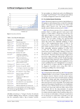
Number of publications 50 38 4.1. Co-citation based clustering
60
40
Figure 3 illustrates the most crucial nodes identified based on
30
the maximum cited references from a total of 15 influential
20
marked with a distinct color. The clusters encapsulate
10 5 9 7 8 4 5 7 6 12 11 15 19 papers. These papers are classified into five clusters, each
11 2 11 1 2 33
0 specific research themes, as summarized in Table 3.
1998 1999 2000 2001 2002 2003 2004 2005 2006 2007 2008 2009 2010 2011 2012 2013 2014 2015 2016 2017 2018 2019 2020 2021 Table 3 provides an overview of the co-citation network
Publication year analysis, where co-citation generally tracks papers cited
Figure 2. Research productivity. together in source articles. Clusters are formed when the
same pair of papers is co-cited by different authors. The
table delineates five clusters, each representing different
Table 1. List of top 20 cited papers
themes. The first cluster (cluster red) predominantly
Reference Notable work LC TC focuses on the AI application in MRIs. Notable references
Litjens et al. [30] Deep learning survey 20 234 within this cluster were uniformly published in either
Wang et al. [31] ML analysis of MR radiomics 15 118 2018 or 2019, emphasizing the impact of MRIs on the
classification or characterization of prostate cancer. The
[32]
Tabesh et al. Multi-feature diagnostics 14 273 green cluster addresses prostate cancer grading systems,
Strom et al. AI model for diagnosis using biopsies 13 112 with noteworthy references published in either 2015
[33]
Chen et al. [34] Radiomic model compared 11 44 or 2016. The blue cluster describes AI-based cancer
to cancer images classification using MRI, sharing a temporal overlap with
Bonekamp et al. [35] Radiomic ML model 11 98 the green cluster as references were published between
Nir et al. [36] Classification of digitized 10 53 2015 and 2017. The yellow cluster discusses AI applications
images with expert learning in prostate cancer pathology, with notable references for
[37]
Gorelick et al. Detection/classification 10 77 this cluster published in 2018 and 2019. Finally, the purple
using prostate histopathology cluster explores deep learning in cancer diagnostics, with
Monaco et al. [38] Detection using probabilistic 10 90 references spanning the largest range compared to all the
pairwise Markov models clusters, published from 2011 through 2018. In conclusion,
Ginsburg et al. Radiomic structures on MRI 8 78 the vast majority of papers describe AI applications in
[39]
Antonelli et al. Prediction through ML classifier 7 31 the diagnosis of prostate cancer, using either MRI or
[40]
Nir et al. [41] AI techniques comparison using images 7 34 pathological grading as key methodologies.
Wang et al. [42] Deep neural network using MRI images 7 63 4.2. Citation-based clustering
[43]
Gertych et al. ML methods for image analyses 7 54 Figure 4 displays the citation network, while Table 4 provides
Tiwari et al. Detection using multi-kernel 7 78 a detailed analysis and summary of themes derived from
[44]
graphing method
Yuan et al. Multiparametric MRI-based 6 38 the citation network. Citation network analysis explores
[45]
classification the specific topics across various research papers, unveiling
Kwak et al. MRI and digital image correlation 6 16 four distinct clusters and their corresponding themes.
[46]
The first cluster (red) describes the AI applications in the
Ozer et al. [47] ML methods for cancer segmentation 6 99 detection of prostate cancer using MRI. The second cluster
Lucas et al. Deep learning techniques 5 44 (green) revolves around AI applications in the classification
[48]
Tiwari et al. [49] Detection using MRI and spectroscopy 5 47 of prostate cancer. The blue cluster focuses on multi-feature
Abbreviations: AI: Artificial intelligence; LC: Location citations; prostate cancer detection, while the yellow cluster delves
ML: Machine learning; MR: Magnetic resonance; MRI: Magnetic into radiomic ML for the characterization of prostate
resonance imaging; TC: Global citations. lesions. It is noteworthy that the four clusters identified in
the citation network analysis differ from those observed in
with links correlating to a ”linkage” of nodes. The network the co-citation network analysis. Although AI applications
is divided utilizing various attitudes, providing insights for the detection and classification of prostate cancer are
into the community within a big co-citation network. common to both co-citation and citation analyses, ML,
Volume 1 Issue 1 (2024) 6 https://doi.org/10.36922/aih.1958