
Page 10 - AIH-2-4
P. 10
Artificial Intelligence in Health AI in acute stroke imaging
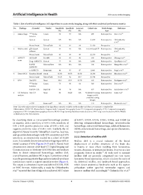
Table 1. List of artificial intelligence (AI) algorithms in acute stroke imaging, along with their analytical performance metrics
No. Findings AI model Vendor Sensitivity Specificity Accuracy Cohort size Study design Reference
(%) (%) (%) type
1 Hemorrhage AUTO Stroke Canon 93 93 NA 200 Retrospective Rava et al. 28
Solution
Qure.ai Qure.ai NA NA NA 21,095 Retrospective Chilamkurthy
et al. 29
Neural Assist TeleradTech 92 84 84 21,420 Prospective -
2 Midline shift qER-Quant Qure.ai 95 95 NA 313,318 head CT Retrospective Chilamkurthy
software et al. 29
Neural Assist TeleradTech 84 89 89 22,729 Prospective -
3 ASPECT AI DLAD D.LABS 65 82 80 258 Retrospective Chiang et al. 43
score Deep-ASPECTS Qure.ai 77 99 NA 5,000 Retrospective Upadhyay et al. 44
RAPID iSchemaView NA NA NA 100 Retrospective Maegerlein et al. 45
ASPECTS
e-ASPECTS Brainomix 44 93 87 2,640 Retrospective Nagel et al. 46
4 Dense MCA Xception Model viso.ai 82.90 89.70 86.50 18,396 Retrospective Shinohara et al. 48
Neural Assist TeleradTech 56.25 94 89.7 22,708 Prospective -
5 LVO Viz-LVO Viz.ai 80.3 82.9 82.70 610 Retrospective Rodrigues et al. 51
AUTO Stroke Canon 73 98 81 303 Retrospective Rava et al. 52
Solution
RAPID-CTA Rapid AI 94 76 NA 477 Retrospective Amukotuwa et al. 53
6 CT Perfusion Viz CTP Viz.ai 80 86.20 NA 94 labeled training Retrospective Soun et al. 1
analysis images and 62
unlabeled testing
images
e-CTP Brainomix ® NA NA NA 111 Retrospective Shahrouki et al. 57
Note: This table enlists selected examples of AI algorithms currently available on the market and does not represent a complete list.
Abbreviations: ASPECTS: Alberta Stroke Program Early Computed Tomography Score; CT: Computed tomography; CTA: Computed tomography
angiograms; CTP: Computed tomography perfusion; DLAD: Deep learning-based automatic detection; LVO: Large vessel occlusion; MCA: Middle
cerebral artery; NA: Not available.
by classifying them as intracranial hemorrhage positive of 0.8977, 0.9559, 0.9194, 0.9161, 0.9044, and 0.9288 for
or negative, with a specificity of 0.93 ± 0.01, sensitivity of detecting intraparenchymal hemorrhage, intraventricular
0.93 ± 0.03, positive predictive value of 0.85 ± 0.02, and hemorrhage, intracranial hemorrhage, subdural hematoma
negative predictive value of 0.98 ± 0.01. Similarly, the AI (SDH), subarachnoid hemorrhage, and epidural hematoma,
algorithm Neural Assist by TeleradTech classifies, localizes, respectively.
and quantifies hemorrhages with 92% sensitivity and 83%
specificity, as prospectively studied in a cohort of 21,420 3.2.2. Detection of midline shift
scans. It accurately detects various hemorrhage types with an Midline shift is a crucial indicator of the lateral
overall accuracy of 85% (Figures 2A-D and 3). Neural Assist displacement of midline structures of the brain due
processes non-contrast adult head CT Digital Imaging and to trauma or mass effects resulting from hematomas,
Communications in Medicine (DICOM) files and analyzes tumors, abscesses, or intracranial lesions. It serves as a key
them to detect intracranial hemorrhage, midline shift, prognostic feature in stroke. AI tools used to measure
37
cranial fractures, and dense MCA signs. It prioritizes critical midline shift are generally categorized into two types:
scans by generating priority flags and automatically produces Symmetry-based approaches, which calculate the curve of
a preliminary report to support specialist review (Figure 4). the deformed midline, and landmark-based approaches,
The output is a structured report available in DICOM, PDF, which detect anatomical landmarks such as the septum
or DOC formats. Additionally, a study by Chilamkurthy pellucidum within specified ventricular regions and
et al. reported that their AI algorithms achieved AUC values measure midline shift accordingly. Chilamkurthy et al.
29
38
29
Volume 2 Issue 4 (2025) 4 doi: 10.36922/AIH025140025