
Page 9 - AIH-2-4
P. 9
Artificial Intelligence in Health AI in acute stroke imaging
were duplicates, conference proceedings, commentaries, strokes due to atherothrombotic disease. Non-invasive
editorials, or abstracts without full-text access. imaging techniques used to assess the likelihood of
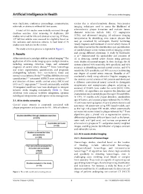
A total of 316 studies were initially retrieved through atherosclerotic plaque formation and evaluate lumen
database searches. After removing 33 duplicates, 283 diameter reduction include MRI, CT angiograms
studies remained for title and abstract screening. Of these, (CTA), and ultrasound imaging. AI enhances imaging
127 full-text articles were assessed for eligibility based on interpretation by identifying even minute plaques that
the inclusion and exclusion criteria. A final total of 78 may go unnoticed by radiologists, thereby facilitating
studies were included in the review. timely diagnosis and treatment of carotid artery disease. It
also helps standardize the identification and quantification
The study selection process is depicted in Figure 1. of carotid plaque across various medical imaging centers
3. Results and among different physicians. A study by Kordzadeh
et al. demonstrated the applicability and precision of
25
AI has introduced a paradigm shift in medical imaging. The AI in detecting carotid artery disease using grayscale
24
application of AI in stroke imaging spans multiple domains, static duplex ultrasound images. In their findings, the AI
including screening, detection, triage, and automated system achieved 91% sensitivity, 86% specificity, and 92%
diagnosis of carotid artery disease, 25-27 brain hemorrhage accuracy in identifying normal carotid arteries, and 87%
and infarct segmentation, quantification, and prognosis; sensitivity, 82% specificity, and 90% accuracy in detecting
distinguishing ischemic from non-ischemic tissue and any degree of carotid artery stenosis. Skandha et al.
26
normal versus infarcted brain; 28-36 midline shift detection and conducted a study using echocolor Doppler imaging on
quantification; 37-41 automated ASPECT score calculation; 42-46 the internal carotid arteries of 345 patients and developed
and detection of dense middle cerebral artery (MCA) and a diffusion convolutional neural network to distinguish
LVO on CT angiograms. 47-53 Several commercially available symptomatic and asymptomatic plaques, achieving an
AI-integrated workflows have been developed to interpret accuracy of 95.66% (area under the curve [AUC] 0.956,
ischemic stroke imaging automatically (Table 1). These p<0.0001). AI algorithms also improve the detection and
AI-driven tools enhance workflow integration, optimize characterization of carotid plaques through CTA and MRA.
radiological interpretation, and improve stroke management. In CTA, AI enables early plaque detection, standardizes
3.1. AI in stroke screening quantification, and assesses plaque vulnerability. In MRA,
AI estimates varying degrees of carotid artery stenosis and
Carotid artery stenosis is commonly associated with automates risk assessment using MRI-based models, such
plaque progression and accounts for 10 – 20% of ischemic as the high-risk plaque MRI model, which automatically
estimates risk scores related to plaque vulnerability. These
27
algorithms play a pivotal role as segmentation systems,
differentiating between different layers (such as the lumen,
outer wall, and lipid core), and various components of
atherosclerotic plaque on T1- and proton density-weighted
images, enabling precise identification of plaque contours
and vulnerable lesions.
3.2. AI in acute stroke imaging
3.2.1. Assessment of hemorrhage
Hemorrhagic strokes, classified based on the location
of bleeding, include subarachnoid hemorrhage,
intraparenchymal hemorrhage, and intraventricular
hemorrhage. AI algorithms have shown high sensitivity
28
and specificity in detecting hemorrhages, even in
challenging cases involving small bleeds or complex
brain anatomy. These tools are capable of segmenting and
quantifying hemorrhages, thereby improving classification
and localization. For instance, a study by Rava et al.
28
demonstrated that the AI could automate the detection and
Figure 1. The article search and screening process triage of patients undergoing non-contrast CT (NCCT)
Volume 2 Issue 4 (2025) 3 doi: 10.36922/AIH025140025