
Page 63 - AN-1-1
P. 63
Advanced Neurologyurology
Advanced Ne T-ASL in etiological diagnosis of multiple infarcts
A C D
B
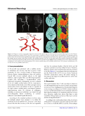
Figure 1. (A) Magnetic resonance angiography image shows absence of the right A1 segment as illustrated in the schematic drawing (B, star). (C) Diffusion-
weighted imaging shows multiple infarcts, which were assumed to be within the territories of the BA and left ICA according to the standard perfusion
territory atlas. (D) Territorial arterial spin labeling perfusion maps show the actual perfusion territories of the right ICA (green), left ICA (purple), and
BA (orange) and the location of the infarct lesions in the perfusion territory of the BA. In this patient with severe stenosis of the left MCA, the flow
territory of the BA had expanded and the perfusion border of the left MCA and left PCA had shifted forward, probably because of the collateral blood flow
compensation. AcoA, anterior communicating artery; ACA, anterior cerebral artery; MCA, middle cerebral artery; PCA, posterior cerebral artery; ICA,
internal carotid artery; and BA, basilar artery
2. Case presentation and that the perfusion border of the left MCA and left
posterior cerebral artery had shifted forward. Therefore,
A 50-year-old man presented with a sudden decline all infarct lesions were located in the perfusion territory
in orientation and memory that had occurred 5 days of the BA (Figure 1). According to the above information
previously. He had a medical history of hypertension, and the Trial of ORG 10172 in Acute Stroke Treatment
diabetes, human immunodeficiency virus, and syphilis. (TOAST) classification criteria, the stroke etiology in
Cranial MRI revealed multiple infarcts in the right this patient was large artery atherosclerosis rather than
midbrain, right occipital lobe, and left temporoparietal- cardiac embolism.
occipital lobe, indicating a thromboembolic event.
Computed tomography angiography revealed occlusion 3. Discussion
of the left middle cerebral artery (MCA), moderate-to- This report illustrates how T-ASL may help to determine
severe stenosis of the basilar artery (BA) and right internal the relationship between the infarct location and perfusion
carotid artery (ICA), and absence of the A1 segment of territories of brain-feeding arteries, thereby identifying the
the right anterior cerebral artery and bilateral posterior stroke etiology according to the TOAST criteria. In clinical
communicating artery. No evidence of cardiogenic practice, the thromboembolic source of multiple infarcts is
embolism or paradoxical embolism was found through determined according to the standard perfusion territory
echocardiography, Holter monitoring, or contrast- atlas. However, studies have shown that there is wide
enhanced transcranial Doppler ultrasound. variability of the perfusion territories of the major cerebral
According to the standard perfusion territory atlas, arteries among individuals [1,2] .
the multiple infarcts were assumed to be within the According to the traditional perfusion atlas, the infarcts
territories of the BA and left ICA. However, T-ASL maps of the patient in the present case were assumed to be within
showed that the flow territory of the BA had expanded the territories of both the left ICA and BA. If the multiple
Volume 1 Issue 1 (2022) 2 https://doi.org/10.36922/an.v1i1.10