
Page 81 - AN-2-1
P. 81
Advanced Ne
Advanced Neurologyurology Choreoathetosis with contralateral substantia nigra infarction
to the patient, grasping the affected limb with his left hand Laboratory examinations showed no abnormalities
prevented the attack. Although there was no disturbance besides hemoglobin A1c and glucose levels. The levels of
of consciousness, slurred speech, clenched teeth, and C-reactive protein, erythrocyte sedimentation rate, liver
salivation at the corners of his mouth, he experienced and renal function, blood lipid, electrolytes, homocysteine,
numbness. The aforementioned symptoms affected his coagulation indices, including antithrombin III, prothrombin
daily life. The patient had no significant medical history. time, activated partial thromboplastin time, and D-dimer,
A physical examination on admission revealed that he had were all within normal limits. Antinuclear antibody, anti-
a temperature of 36.4°C, a heart rate of 80 beats/min, a dsDNA antibody, anti-Smith (Sm) antibody, anti-SS-A/B
respiratory rate of 18 breaths/min, and a blood pressure antibody, proteinase 3 antineutrophil cytoplasmic antibody
reading of 140/80 mmHg. Cardiopulmonary and (PR3-ANCA), myeloperoxidase antineutrophil cytoplasmic
abdominal examination showed no obvious abnormalities. antibody (MPO-ANCA), as well as beta-2-glycoprotein I,
Neurological examination indicated that he was fully human immunodeficiency virus (HIV), and syphilis
conscious and oriented. Bilateral pupils, about 3 mm antibodies were all negative. The laboratory results
in diameter, were sensitive to light reflex. Except for the are shown in Table 1. The patient was diagnosed with
involuntary dance-like movements of the right upper limb, contralateral SN infarction and was started on antiplatelet
there were no positive signs. therapy with aspirin 100 mg and clopidogrel 75 mg orally
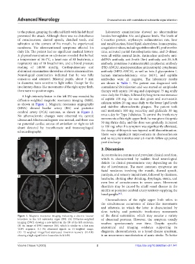
A high-intensity lesion in the left SN was revealed by once daily for 21 days, followed by long-term maintenance
diffusion-weighted magnetic resonance imaging (MRI), of aspirin 100 mg. He was also given oral atorvastatin
as shown in Figure 1. Magnetic resonance angiography calcium tablets 20 mg once daily to the lower lipid levels
(MRA) showed basilar artery (BA) and posterior and stabilize atherosclerotic plaques. The patient took
cerebral artery (PCA) stenoses, as shown in Figure 2. oral metformin 500 mg twice/day and gliclazide 30 mg
No atherosclerotic changes were observed via carotid once a day for Type 2 diabetes. To control the involuntary
ultrasound. Electrocardiogram was normal, and there was movements of his right upper limb, he was given thiopride
no potential cardiac source of embolism or right-to-left 50 mg thrice daily, and the dose was gradually increased
shunt detected by transthoracic and transesophageal to 100 mg; when his symptom was significantly alleviated,
echocardiography. the dosage of thiopride was tapered until discontinuation.
There were significant improvements in choreoathetosis
A B and no recurrent strokes over a 6-month follow-up period
post-discharge.
3. Discussion
Acute stroke is a common and prevalent clinical condition,
which is characterized by sudden focal neurological
deficit. Its clinical presentations vary depending on the
site of involvement. The most common symptoms are
facial weakness involving the mouth, slurred speech,
paralysis, and sensory impairment, followed by dizziness,
C D
headache, choking after drinking, dysphagia, ataxia, and
even loss of consciousness in severe cases. Movement
disorders may be caused by small vessel disease in the
middle or posterior cerebral artery territory supplying the
basal ganglia [4,5] .
Choreoathetosis of the right upper limb refers to
the simultaneous occurrence of dance-like movements
and athetosis, in which the latter is characterized by
slow, twitchy, and peristaltic involuntary movements
Figure 1. Magnetic resonance imaging indicating a discrete lacunar of the distal extremities, which may assume a variety
infarction in the left substantia nigra (SN). (A) Diffusion-weighted of abnormal postures. However, the symptom usually
imaging (DWI) showing a new infarct in the SN of the left cerebrum. resolves spontaneously over time. Despite specific
(B) An image of DWI sequence (b0), which is similar to a fast-scan anatomical and imaging evidence supporting its
T2WI sequence. (C) No abnormal signals in T1-weighted image.
(D) T2-weighted image/fluid-attenuated inversion-recovery (FLAIR) diagnosis, choreoathetosis, as a broad disease spectrum,
showing a high-signal lesion located in the left SN. is an uncommon manifestation of acute stroke. To better
Volume 2 Issue 1 (2023) 2 https://doi.org/10.36922/an.v2i1.141