
Page 82 - AN-2-1
P. 82
Advanced Neurology Choreoathetosis with contralateral substantia nigra infarction
A B C
D E F
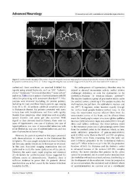
Figure 2. Cerebrovascular imaging of the patient. (A and B) Magnetic resonance angiography revealing atherosclerotic stenosis of the basilar artery and the
left posterior cerebral artery (PCA). (C–F) Four images showing the cross-sectional images of BCA and PCA, with observable left PCA stenosis.
understand these conditions, we searched PubMed for The pathogenesis of hyperactivity disorders may be
reports using several keywords, such as “SN,” “ischemic related to elevated monoamine activity and/or relative
stroke,” “dyskinesia,” “movement disorders,” “motor deficit,” cholinergic deficiency, or even the dysfunction in the
and so on. Table 2 is a summary of several patients with SN cerebellum-thalamic or striatum-thalamic pathway .
[8]
infarction presenting with movement disorders [6-14] . Nine The nucleus basalis is a group of grey matter nuclei under
patients were reviewed (excluding the present patient), the cerebral cortex, consisting of the caudate nucleus, the
including six male and three female patients, age ranging shell nucleus, the pallidum, the subthalamic nucleus, and
from 18 to 83. All patients exhibited symptoms similar the SN . It regulates motor function mainly through
[15]
to Parkinson’s disease. Six patients presented with static the cortico-basal ganglia-thalamo-cortical loop. In this
tremor, four with bradykinesia, and three with rigidity. loop, the neostriatum receives afferent fibers from the
Besides these symptoms, other symptoms such as apathy, sensorimotor cortex of the brain, and its efferent fibers
speech disorder, and panic gait also occurred. With reach the basal ganglia output structures (globus pallidus
regard to their previous medical history, there were six internus [GPi]/substantia nigra reticularis [SNr]) through
cases of hypertension, one case of diabetes, one case of direct and indirect pathways. The direct pathway refers
hyperlipidemia, one case of myocardial infarction with to the excitatory projection of glutaminergic nerve fibers
atrial fibrillation, one case of cerebral infarction, and one from the cerebral cortex to the striatum, which, in turn,
case of subarachnoid hemorrhage. sends inhibitory projections of gamma-aminobutyric
However, the patient reported in this paper presented acid (GABA) nerve fibers to the GPi and SNr complexes.
with choreoathetosis in contrast to the Parkinson’s-like The GABA nerve fibers emanating from the latter can
symptoms. This case caught out attention since there have emit inhibitory projection to the ventrolateral thalamic
[16]
not been any similar reports in the literature we reviewed. nucleus . When infarcts occur in the SN, the projection
In this case, functional imaging examination was not function of both the thalamus and the thalamic cortex
performed to verify the motor dysfunction of the patient. is enhanced because of the reduced GPi-SNr complex
However, the acute onset, rapid progress, and existence activity. The motor impulses that generate in the cortex
of risk factors for cerebrovascular disease suggested the cannot be terminated, and it cannot receive impulses from
different causes of choreoathetosis. Therefore, there is the cerebellar-red nucleus-striatal pathway, thus leading to
sufficient evidence to prove that the patient’s clinical the development of choreoathetosis. From the perspective
manifestation was in fact caused by the lacunar infarction of neurotransmitters, the excitatory and inhibitory effects
located in the SN. of neurotransmitters on neurons are in a coordinated
Volume 2 Issue 1 (2023) 3 https://doi.org/10.36922/an.v2i1.141