
Page 112 - AN-3-1
P. 112
Advanced Neurology MNS in patients with anxiety and depressive symptoms
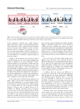
Figure 3. Scheme for obtaining results: Mirror tasks caused clear mu-rhythm suppression in the central regions of the brain in the upper-frequency range
(10.5 – 13 Hz) in the control group. Patients showed suppression of EEG rhythms over a wider range of frequencies and in several areas of the brain and
the greatest activation of the MNS was in the tasks of observation and joint execution of hand clenching.
Second, suppression occurred over a wider frequency may mean that patients suffering from affective disorders
range in the alpha-1, alpha-2, and beta frequency bands. engage in additional resources and neural networks to
Third, suppression mainly occurred during observation complete the tasks. Available data support the notion
and joint execution tasks. Notably, the level of rhythm that patients with anxiety and depressive symptoms have
desynchronization in patients was generally not lower weak and inefficient brain networks and require additional
than that in the healthy subjects. Thus, no insufficiency or neural resources. 33,45 This also indicates that the MNS is
decrease in the activity of the system was established, and a functional network that is not tightly bound to specific
there were differences in localization, frequency band, and brain structures and may involve additional non-specific
selectivity in tasks. areas.
Changes in the localization and a frequency shift of The mirror system of the human brain contains many
functional rhythms are the most common findings from different areas. The MNS core is generally thought to
studies on depressive and anxiety disorders. Spatial- include the following cortical regions: the inferior parietal
51
frequency changes in the organization of brain activity in lobule, especially the supramarginal gyrus and adjacent
patients with anxiety-affective disorders have also been intraparietal sulcus, ventral premotor cortex, and posterior
confirmed by other authors. 52-57 Thus, if the frequency of inferior frontal gyrus. However, the extended MNS
the main rhythm is shifted in patients as a result of their contains additional regions, including the primary motor
mental state, eventhough it remains functionally similar to cortex and supplementary motor area. 13,58
the alpha or mu rhythm, the suppression of the alpha or In addition, there are features and differences in
mu rhythm would appear in a wider range of frequencies. the areas and connections of the MNS that implement
Therefore, a shift in basic functional EEG rhythms may hand movements and facial expressions. Difficulties
59
reflect a change in the functional state of the brain. with identifying specific structures also lie in the fact
In this study, patients exhibited strong suppression of that it is impossible to clearly limit the functions of
alpha-1, alpha-2, and even beta-1 rhythms in the occipital, the MNS to: projection of movements, projection and
central, and frontal regions. It can be assumed that this shift emotional assessment, projection, emotional assessment,
in the frequency and spatial distribution reflects changes and understanding of the goal, or understanding the
in the extended MNS and other associated functional motive of the opponent’s actions. Thus, spatial activation
networks. Activation areas in patients defined by the when observing and performing movements can vary
sLORETA are diffusely distributed almost throughout significantly and can include many different areas.
the entire volume of the brain, affecting both the frontal Numerous studies have identified that the human
lobes and visual areas, with the highest intensity recorded mirror system is, to some extent, both a product and a
in the alpha-1 range. Significant intensity in patients was process of social interaction. It has been shown that it is
60
registered in Brodmann areas 8 and 9 of the frontal lobes, possible to retrain the MNS with motor training. These
61
in the associative visual field 19 and fields 35 and 36. This results indicate that the mirror properties of the mirror
Volume 3 Issue 1 (2024) 8 https://doi.org/10.36922/an.2009