
Page 129 - AN-3-1
P. 129
Advanced Neurology Seizure as the first symptom of CS-DAVF
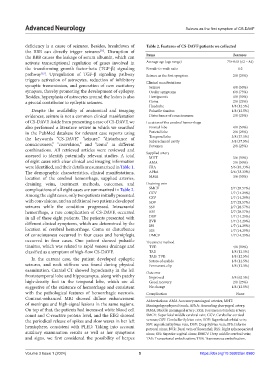
deficiency is a cause of seizures. Besides, breakdown of Table 2. Features of CS‑DAVF patients we collected
the BBB can directly trigger seizures . Disruption of
[11]
the BBB causes the leakage of serum albumin, which can Items Features
activate transcriptional regulation of genes involved in Average age (age range) 75±8.65 (62 – 84)
the transforming growth factor-beta (TGF-β) signaling Female-to-male ratio 6:2
pathway . Upregulation of TGF-β signaling pathway Seizure as the first symptom 2/8 (25%)
[12]
triggers activation of astrocytes, reduction of inhibitory Clinical manifestations
synaptic transmission, and generation of new excitatory Seizure 4/8 (50%)
synapses, thereby promoting the development of epilepsy. Ocular symptoms 6/8 (75%)
Besides, hyperplasia of astrocytes around the lesion is also Hemiparesis 4/8 (50%)
a pivotal contributor to epileptic seizures. Coma 2/8 (25%)
Headache 1/8 (12.5%)
Despite the availability of anatomical and imaging Pulsatile tinnitus 1/8 (12.5%)
evidences, seizure is not a common clinical manifestation Disturbance of consciousness 2/8 (25%)
of CS-DAVF. Aside from presenting a case of CS-DAVF, we Location of the cerebral hemorrhage
also performed a literature review in which we searched Frontal lobe 4/8 (50%)
in the PubMed database for relevant case reports using Parietal lobe 2/8 (25%)
the keywords “CS-DAVF,” “seizure,” “disturbance of Temporal lobe 3/8 (37.5%)
Subarachnoid cavity
3/8 (37.5%)
consciousness,” “convulsion,” and “coma” in different Putamen 2/8 (25%)
combinations. All retrieved articles were reviewed and
assessed to identify potentially relevant studies. A total Supplied artery 3/6 (50%)
MHT
of eight cases with clear clinical and imaging information AMA 3/6 (50%)
were identified, and their details are summarized in Table 1. FRA 5/6 (83.33%)
The demographic characteristics, clinical manifestations, APhA 2/6 (33.33%)
location of the cerebral hemorrhage, supplied arteries, MMA 3/6 (50%)
draining veins, treatment methods, outcomes, and Draining vein
complications of all eight cases are summarized in Table 2. SMCV 2/7 (28.57%)
Among the eight cases, only two patients initially presented CCV 1/7 (14.29%)
CSV
1/7 (14.29%)
with convulsions, and an additional two patients developed SOV 2/7 (28.57%)
seizures while the condition progressed. Intracranial SSV 2/7 (28.57%)
hemorrhage, a rare complication of CS-DAVF, occurred RSS 2/7 (28.57%)
in all of these eight patients. The patients presented with DSV 1/7 (14.29%)
different clinical symptoms, which are determined by the BVR 1/7 (14.29%)
1/7 (14.29%)
IPS
location of cerebral hemorrhage. Coma or disturbance SSS 1/7 (14.29%)
of consciousness occurred in four cases and hemiplegia DMCV 1/7 (14.29%)
occurred in four cases. One patient showed pulsatile Treatment method
tinnitus, which was related to rapid venous drainage and TVE 4/8 (50%)
classified as a symptom of high-flow CS-DAVF. TAE 1/8 (12.5%)
TAE+TVE 1/8 (12.5%)
In the current case, the patient developed epileptic Suture of eyelids 1/8 (12.5%)
seizures, and neck stiffness was found during physical Permanent clip 1/8 (12.5%)
examination. Cranial CT showed hypodensity in the left Outcome
frontotemporal lobe and hippocampus, along with patchy Improved 5/8 (62.5%)
high-density foci in the temporal lobe, which are all Good recovery 2/8 (25%)
suggestive of the existence of hemorrhage and consistent No change 1/8 (12.5%)
with the pathological features of hemorrhagic necrosis. Complication None
Contrast-enhanced MRI showed diffuse enhancement Abbreviations: AMA Accessory meningeal arteries; MHT:
of meninges and high-signal lesions in the same regions. Meningohypophyseal trunk; APhA: Ascending pharyngeal artery;
On top of that, the patients had increased white blood cell MMA: Middle meningeal artery; FRA: Foramen rotundum artery;
count and C-reactive protein level, and the EEG showed SMCV: Superficial middle cerebral vein; CCV: Cerebellar cortical
the periodical release of spikes and slow waves in her left venous; CSV: Cerebellar Sylvian vein; SOV: Superficial orbital vein;
hemisphere, consistent with PLED. Taking into account SSV: superficial Sylvian vein; DSV: Deep Sylvian vein; IPS: Inferior
petrosal sinus; BVR: Basal vein of Rosenthal; RSS: Right sphenoparietal
auxiliary examination results as well as her symptoms sinus; SSS: Superior sagittal sinus; DMCV: Deep middle cerebral vein;
and signs, we first considered the possibility of herpes TAE: Transarterial embolization; TVE: Transvenous embolization.
Volume 3 Issue 1 (2024) 5 https://doi.org/10.36922/an.0980