
Page 126 - AN-3-1
P. 126
Advanced Neurologyurology
Advanced Ne Seizure as the first symptom of CS-DAVF
before the onset of invasive symptoms, probably related to patchy hemorrhage in the left temporal lobe, and dilatation
multiple shunts of blood flow , and seizures only occurred of the left superior ophthalmic vein (SOV) (Figure 1). Brain
[2]
in a minority of patients with CS-DAVF . Here, we report scan and contrast-enhanced magnetic resonance imaging
[3]
an unusual case of CS-DAVF presenting with seizures (MRI) showed diffuse meningeal enhancement and high
as the first symptom, focusing on the diagnosis, clinical signal changes in the left frontoparietal temporal lobe,
course, and treatment outcome. insular lobe, and hippocampal head region (Figure 2).
Electroencephalography (EEG) showed the periodical
2. Case presentation release of spikes and slow waves in the left hemisphere,
A 62-year-old Chinese woman with paroxysmal loss of consistent with periodic lateralized epileptiform
consciousness, involuntary eye movement to the left, discharges (PLED) (Figure 3). The patient underwent total
spasm of the right facial muscles, chewing issues, salivation cerebral angiography under general anesthesia (Figure 4)
at the right corner of the mouth, and bit tongue was after giving the informed consent. Digital subtraction
admitted to our hospital. The patient did not complain of angiography revealed that the CS-DAVF arterial supply
convulsions or limb clonus, urinary incontinence, and fecal originated in the dural branch of the left external carotid
incontinence. The patient had gone through three episodes artery (ECA), indicating a condition classified as type C
of loss of consciousness, and every time after she regained under the barrow caroticocavernous fistula classification,
consciousness, her response lasted very briefly. After with venous drainage into the ipsilateral cavernous sinus,
wide-awake, she was able to understand others’ words and intercavernous sinus, contralateral cavernous sinus, left
respond in a simple manner, with three episodes. However, SOV, left lateral fissure vein, and left pterygoid venous
on the 2 day of admission, the patient’s response became plexus. Accordingly, transvenous embolization was
nd
weaker than before, with intermittent involuntary grip.
The patient had a previous medical history of breast cancer
and ulcerative colitis, without hypertension, diabetes,
and coronary heart disease. Almost none of her family
members require any medical attention, except for her
mother who had type 2 diabetes.
Physical examination at admission revealed that she had
a body temperature of 36.7°C, a heart rate of 101 bpm, a
respiratory rate of 20 breaths/min, and a blood pressure
measurement of 168/95 mmHg. We did not uncover any
obvious abnormalities from the cardiopulmonary and
abdominal examination. Neurologic examination indicated
that she was in a state of somnolence. The neck was slightly
tonic. However, the patient was not being cooperative to
complete the rest of the neurological physical examinations.
The blood test results revealed high white cell count at Figure 1. Computed tomography scan showing hemorrhage in the
14.69 × 10 /L (reference: 3.5 – 9.5 × 10 /L), high level of left temporal lobe, edema in the left frontotemporal lobe and basal
9
9
ganglia, bulging of cavernous sinus, and distension of the left superior
C-reactive protein at 47.98 mg/L (reference: 0 – 10 mg/L), and ophthalmic vein.
high level of procalcitonin at 0.445 ng/mL (<0.046 ng/mL).
The pressure of cerebrospinal fluid (CSF) was 190 mm H O. A B
2
Results of the CSF routine and biochemical tests are as
follows: Glucose 4.84 mmol/L, chlorine ion 114.4 mmol/L,
protein 0.41 g/L, CSF-IgG 50.2 g/L, CSF-IgA 6.4 g/L, and
CSF-IgM 1.5 g/L. Antibody titer of a few viruses, including
rubella virus, cytomegalovirus, herpes simplex virus, in
CSF was all within the normal limits. Aside from that,
negative results were noted in tests for DNA and antibody
of Mycobacterium tuberculosis, fungi, and autoimmune
encephalitis antibody markers in CSF.
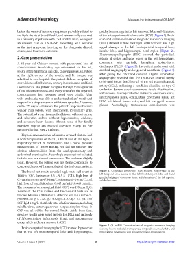
Figure 2. (A and B) Contrast-enhanced magnetic resonance imaging
Brain computed tomography (CT) showed hypodense showing lesions in the left frontoparietal temporal lobe, insular lobe, and
foci in the left frontotemporal lobe and hippocampus, hippocampal head region with diffuse meningeal enhancement.
Volume 3 Issue 1 (2024) 2 https://doi.org/10.36922/an.0980