
Page 128 - AN-3-1
P. 128
Advanced Neurology Seizure as the first symptom of CS-DAVF
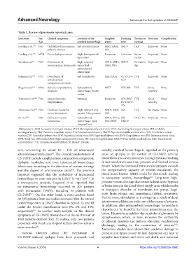
Table 1. Review of previously reported cases.
Literature Sex/ Clinical symptoms Location of the Supplied Draining Treatment Outcome Complication
Age cerebral hemorrhage artery vein method
Harding et al. [13] F/63 Ophthalmodynia; seizure; Left precentral gyrus MHT; AMA; SMCV TAE Improved None
pulsatile tinnitus FRA
Harding et al. [13] M/74 Ptosis; diplopia; seizure Right frontoparietal Unknown Unknown Suture Improved None
operculum eyelids
Tanioka et al. [2] F/80 Disturbance of Right temporal APhA; MMA; SMCV Permanent Improved None
consciousness; hemiparesis subcortical; AMA; FRA clip
subarachnoid
hemorrhage
Sakuma et al. [14] F/73 Disturbance of Left frontal lobe FRA; RICA CCV; CSV; TVE Improved None
consciousness; SOV
hemiparesis; chemosis
Meguro et al. [15] M/62 Seizure; exophthalmos; Subarachnoid MHT SSV; RSS TVE Good None
chemosis hemorrhage; frontal recovery
cortex
Takazawa et al. [16] F/81 Seizure; hemosis; Putamen Unknown SSV; DSV; TVE Good None
exophthalmos BVR; SOV; recovery
IPS
Akamatsu et al. [17] F/84 Chemosis; headache; Right temporal and MMA; AMA; RSS TVE No change None
coma; hemiparesis parietal lobe; putamen FRA
Iki et al. [3] F/83 Coma; anisocoria; Subarachnoid MMA; APhA; SSS; TAE; TVE Improved None
hemiparesis hemorrhage; right FRA; MHT DMCV
temporal lobe
Abbreviations: AMA Accessory meningeal arteries; MHT: Meningohypophyseal trunk; APhA: Ascending pharyngeal artery; MMA: Middle
meningeal artery; FRA: Foramen rotundum artery; ICA: Internal carotid artery; SMCV: Superficial middle cerebral vein; CCV: Cerebellar cortical
venous; CSV: Cerebellar Sylvian vein; SOV: Superficial orbital vein; SSV: Superficial Sylvian vein; DSV: Deep Sylvian vein; IPS: Inferior petrosal sinus;
BVR: Basal vein of Rosenthal; RSS: Right sphenoparietal sinus; SSS: Superior sagittal sinus; DMCV: Deep middle cerebral vein; TAE: Transarterial
embolization; TVE: Transvenous embolization, M: Male, F: Female.
vein, accounting for about 10 – 15% of intracranial notably, cerebral hemorrhage is regarded as the primary
arteriovenous fistula cases . The clinical manifestations of cause of epilepsy in the context of CS-DAVF. Arterial
[4]
CS-DAVF include exophthalmos, conjunctival congestion, blood flows into cavernous sinus through fistulas, resulting
diplopia, headache, and even intracranial hemorrhage, in increased cavernous sinus pressure and blocked venous
which vary according to the direction of venous drainage return. When the increased intravascular pressure exceeds
and the degree of arteriovenous shunt . The previous the compensatory capacity of venous circulation, the
[5]
literature suggested that the probability of intracranial blood-brain barrier (BBB) could be destroyed, leading
[6]
hemorrhage or even seizures in DAVF is very low . In to secondary cerebral hemorrhage . Long-term high-
[6]
a retrospective analysis, Cognard et al. reported that pressure venous drainage also causes ischemia and hypoxia
no intracranial hemorrhage occurred in 205 patients of brain tissue in the distal blood supply area, which results
with intracranial DAVFs, including 33 patients with in transport disorder of membrane ion pump, large-
[7]
CS-DAVF . On the other hand, a meta-analysis based scale brain edema, and neurological deficit symptoms.
on 395 patients from six studies estimated that the annual Furthermore, stimulation to meninges as a result of dural
hemorrhage rates in DAVF classified as types I, II, and III arteriovenous dilatation is also one of the causes of seizures.
under the Borden classification were 0%, 6%, and 10%, In addition, after intracerebral hemorrhage, hemosiderin
respectively . In another meta-analysis on the clinical deposits can be found in the brain parenchyma near the
[8]
symptoms of CS-DAVF, Alexandre et al. found that out of lesion. Hemosiderin inhibits the reuptake of glutamate by
1043 patients derived from 22 studies, only two patients synaptosomes, which, in turn, increases the excitability
presented with focal neurological deficits and no seizures of adjacent neurons and induces abnormal synchronous
discharge, leading to the occurrence of seizures .
[10]
were recorded .
[9]
Numerous studies have shown that oxidative damage to
Various theories about the mechanism of proteins and lipids caused by iron deposition can lead to
CS-DAVF-induced epilepsy have been proposed, and synaptic inactivation and nerve cell death, and neuronal
Volume 3 Issue 1 (2024) 4 https://doi.org/10.36922/an.0980